Recent brain imaging studies using functional magnetic resonance imaging (fMRI) have implicated insula and anterior cingulate cortices in the empathic response to another's pain. However, virtually nothing is known about the impact of the voluntary generation of compassion on this network. To investigate these questions we assessed brain activity using fMRI while novice and expert meditation practitioners generated a loving-kindness-compassion meditation state. To probe affective reactivity, we presented emotional and neutral sounds during the meditation and comparison periods. Our main hypothesis was that the concern for others cultivated during this form of meditation enhances affective processing, in particular in response to sounds of distress, and that this response to emotional sounds is modulated by the degree of meditation training. The presentation of the emotional sounds was associated with increased pupil diameter and activation of limbic regions (insula and cingulate cortices) during meditation (versus rest). During meditation, activation in insula was greater during presentation of negative sounds than positive or neutral sounds in expert than it was in novice meditators. The strength of activation in insula was also associated with self-reported intensity of the meditation for both groups. These results support the role of the limbic circuitry in emotion sharing. The comparison between meditation vs. rest states between experts and novices also showed increased activation in amygdala, right temporo-parietal junction (TPJ), and right posterior superior temporal sulcus (pSTS) in response to all sounds, suggesting, greater detection of the emotional sounds, and enhanced mentation in response to emotional human vocalizations for experts than novices during meditation. Together these data indicate that the mental expertise to cultivate positive emotion alters the activation of circuitries previously linked to empathy and theory of mind in response to emotional stimuli.
(AI) and (Ins.) stand for anterior insula and insula, respectively (z = 12 and z = 19, 15 experts and 15 novices, color codes: orange, p less than 5.10ˆ-2, yellow, p less than 2.10ˆ-2). B, C. Impulse response from rest to compassion in response to emotional sounds in AI (B) and Ins. (C). D–E. Responses in AI (D) and Ins. (E) during poor and good blocks of compassion, as verbally reported, for 12 experts (red) and 10 novices (blue).
Monday, March 31, 2008
Regulating the brain circuits of compassion
Here is yet more compelling evidence that you are what you spend your time imagining. In a recent study in PLoS ONE, Lutz, Davidson and colleagues extend their previous work on correlations between brain states and meditation to show that one particular kind of Buddhist meditation, which emphasizes empathetic and loving thoughts towards others and self, changes the brain's reactivity to emotional sounds. In experienced practitioners of the 'loving-kindness-compassion' meditation technique, such images caused larger reactions in the insular and anterior cingulate cortices than were observed in novices. Here is their abstract and one figure from the paper.

Sunday, March 30, 2008

Friday, March 28, 2008
Rodent trained to be Las Vegas croupier
Atshushi Iriki's group in Tokyo has trained degus (intelligent rodents native to the highlands of Chile) to provide the first example (published in PLOS ONE) of rodents wielding tools for a task. (see my 5/7/2007 post for an example with Ravens. Monkeys and Chimps also use tools - Hihara et al. have found extension of corticocortical afferents into the anterior bank of the intraparietal sulcus after tool-use training in adult monkeys.) It will be interesting to see whether tool-use training in degus also results in extended representations in parietotemporal areas and newly formed connections between brain areas, including the prefrontal cortex, similar to those observed in the macaque brain. Work of this sort begins to define brain structures used in the development of tool use.
The mind's eye in number space
From Loetscher et al., an interesting bit on how our subtle muscle movements correlate with counting operations - numbers and space:
Human subjects' answer to questions like “what number is halfway between 2 and 8” provides insights into spatial attention mechanisms involved in numerical processing. Here we show that mental numerical bisections are accompanied by a systematic pattern of horizontal eye movements: processing of a large number followed by a small number is accompanied with leftward eye movements, a tendency less pronounced or even reversed for the processing of a small number followed by a large number. The eyes thus appear to move along a left-to-right-oriented number line, indicating that shifts of attention in representational space are accompanied by an ocular motor orienting response. These results add to the growing evidence for a convergence of numerical processing, spatial attention, and movement planning in the parietal and frontal lobes. They also demonstrate the homologous relationship between our internal representations of numbers and space, and show that the concept of “number space” is more than a mere metaphor.
Thursday, March 27, 2008
A hierarchy of temporal receptive windows in our brains
Here is the abstract from a fascinating study by Hasson et al. on how our visual system assembles time narratives - as during watching a movie - followed by part of one of the figures from the paper:
Real-world events unfold at different time scales and, therefore, cognitive and neuronal processes must likewise occur at different time scales. We present a novel procedure that identifies brain regions responsive to sensory information accumulated over different time scales. We measured functional magnetic resonance imaging activity while observers viewed silent films presented forward, backward, or piecewise-scrambled in time. Early visual areas (e.g., primary visual cortex and the motion-sensitive area MT+) exhibited high response reliability regardless of disruptions in temporal structure. In contrast, the reliability of responses in several higher brain areas, including the superior temporal sulcus (STS), precuneus, posterior lateral sulcus (LS), temporal parietal junction (TPJ), and frontal eye field (FEF), was affected by information accumulated over longer time scales. These regions showed highly reproducible responses for repeated forward, but not for backward or piecewise-scrambled presentations. Moreover, these regions exhibited marked differences in temporal characteristics, with LS, TPJ, and FEF responses depending on information accumulated over longer durations (~36 s) than STS and precuneus (~12 s). We conclude that, similar to the known cortical hierarchy of spatial receptive fields, there is a hierarchy of progressively longer temporal receptive windows in the human brain.
Figure- Maps are shown on inflated (top) and unfolded (bottom) left and right hemispheres. White outlines mark the main regions in which responses were not time reversible. Anatomical abbreviations: ITS, inferior temporal sulcus; LS, lateral sulcus; STS, superior temporal sulcus; TPJ, temporal parietal junction; CS, central sulcus; IPS, intraparietal sulcus. Several higher-order visual areas were functionally defined based on their responses to faces (red outlines), objects (blue outlines), and houses (green outlines). Functionally and anatomically defined cortical areas: V1, primary visual cortex; MT+, MT complex responsive to visual motion; PPA, parahippocampal place area; FFA, fusiform face area; LO, lateral occipital complex responsive to pictures of objects; STS-face, area in superior temporal sulcus responsive to faces.

Wednesday, March 26, 2008
Anxiety: fear in seach of a cause
 The title of this post is a pithy definition that Patricia Pearson gives in her recent book, "A BRIEF HISTORY OF ANXIETY... Yours and Mine", reviewed by William Grimes in the NY Times. From that review some clips:
Everywhere and nowhere, anxiety... In many cases it is the fear of fear itself, a free-floating, nebulous entity that, like a mutant virus, feeds on any available host. Reason is powerless against it. Ms. Pearson argues, in fact, that rationalism, intended to banish superstition and fear, has instead removed one of the most effective weapons against anxiety, namely religious faith and ritual.
...the worship of reason and science, by encouraging the notion that human beings can control their environment, has created a terrible fault line in the modern psyche, although not all societies suffer equally. Mexicans have lots to worry about but don’t. The World Mental Health Survey, conducted in 2002, found that only 6.6 percent of Mexicans had ever experienced a major episode of anxiety or depression. Meanwhile, to their north, 28.8 percent of the American population has been afflicted with anxiety, the highest level in the world. Mexicans who move to the United States adapt, becoming more anxious.
Depressing news: antidepressants don't work?
In the April issue of Nature Reviews Neuroscience, Claudia Wiedemann reviews reactions to a meta analysis by Kirsch et al. of data on antidepressant drugs submitted to the Food and Drug Administration that resulted in the licensing of four of the most commonly prescribed antidepressants: the selective serotonin reuptake inhibitors (SSRIs) Prozac, Seroxat, Effexor and Serzone. For anything but the most severe depression, there was no difference between the drugs and placebos. Kirsch suggests that there is little reason to prescribe anti-depressant medication to any but the most severely depressed patients. The conclusion of the study:
Drug–placebo differences in antidepressant efficacy increase as a function of baseline severity, but are relatively small even for severely depressed patients. The relationship between initial severity and antidepressant efficacy is attributable to decreased responsiveness to placebo among very severely depressed patients, rather than to increased responsiveness to medication.
Tuesday, March 25, 2008
Differing perception of facial expressions in the East and West
 Nagourney describes a study in the March issue of The Journal of Personality and Social Psychology reinforcing previous work showing that Westerners are more likely to see emotions as individual feelings while East Asians see them as inseparable from the feelings of the group. Many researchers have suggested that East Asians take a more holistic view of the world. Here is the abstract of the Masuda et al. article:
Two studies tested the hypothesis that in judging people's emotions from their facial expressions, Japanese, more than Westerners, incorporate information from the social context. In Study 1, participants viewed cartoons depicting a happy, sad, angry, or neutral person surrounded by other people expressing the same emotion as the central person or a different one. The surrounding people's emotions influenced Japanese but not Westerners' perceptions of the central person. These differences reflect differences in attention, as indicated by eye-tracking data (Study 2): Japanese looked at the surrounding people more than did Westerners. Previous findings on East-West differences in contextual sensitivity generalize to social contexts, suggesting that Westerners see emotions as individual feelings, whereas Japanese see them as inseparable from the feelings of the group.
Heritability of cooperative behavior
A study of the behaviors of monozygotic versis dizygotic twins (i.e. 'identical' vs. 'non-identical' twins) in a classical cooperation game yields evidence for genetic influences on yet another of our behaviors - how trusting we are:
Although laboratory experiments document cooperative behavior in humans, little is known about the extent to which individual differences in cooperativeness result from genetic and environmental variation. In this article, we report the results of two independently conceived and executed studies of monozygotic and dizygotic twins, one in Sweden and one in the United States. The results from these studies suggest that humans are endowed with genetic variation that influences the decision to invest, and to reciprocate investment, in the classic trust game. Based on these findings, we urge social scientists to take seriously the idea that differences in peer and parental socialization are not the only forces that influence variation in cooperative behavior.
Our results are complementary to work on the neurological and hormonal substrates of behavior in the trust game and other similar social dilemma games...Enhanced oxytocin levels have been documented in subjects who received a monetary transfer that reflected an intention of trust, and later work has demonstrated that exogenously administered oxytocin increases trust. Scholars have also documented associations between cortisol and trust. These hormonal studies, therefore, indicate that further study of polymorphisms of CYP11B1, OXTR, and other genes involved in the expression and regulation of these hormones may explain part of the genetic effect on cooperation. In fact, one research team has already identified a polymorphism in the AVPR1a gene that is associated with related behavior in the dictator game.
Monday, March 24, 2008
Neuroimaging shows use of self thoughts to infer others' mental states
Jenkins et al. offer a fascinating study of how we infer the mental states of others (mentalize), making use of a phenomenon (repetition suppression) that I had not been aware of before. Here I've done a cut/paste/edit from the abstract and article to try to outline the basic idea, and also show the central figure:
One useful strategy for inferring others' mental states (i.e., mentalizing) may be to use one's own thoughts, feelings, and desires as a proxy for those of other people (This approach to social cognition is alternately described as "simulationist," "projectionist," or "self-referential".) A dorsal aspect of the medial prefrontal cortex has been associated with mentalizing about people perceived to be dissimilar from oneself, whereas a more ventral aspect of medial prefrontal cortex (vMPFC) has been linked to mentalizing about those perceived to be similar. Critically, this vMPFC region also has been observed repeatedly during tasks that require participants to introspect about their own mental experiences, suggesting a connection between tasks that require self-referential thought and those that require inferences about the mental states of similar others.
Because such techniques integrate neural activity across hundreds of thousands of neurons, activation of the same brain voxel by different tasks might occur because each activates distinct, but neighboring or interdigitated, neuronal populations. In this way, two tasks could possibly coactivate the same brain voxel despite engaging different sets of neurons that subserve disparate cognitive processes.
This technical limit can now be circumvented by recently developed paradigms that support stronger conclusions regarding the coactivation of the same neurons by different stimuli or different tasks. These techniques rely on an effect known as "repetition suppression," the observation that neural activity in stimulus-sensitive brain regions is typically reduced when a stimulus is repeated
Suppression across two stimuli indicates that the same (or at least a largely overlapping) population of neurons is engaged by both stimuli. For example, a demonstration of repetition suppression for the number "3" when it follows "4" but not when it follows "40" might suggest that a relatively high proportion of the neurons that code for the number "3" also participate in representations of similar numerosities (such as "4"), but not in representations of more distant numerosities.
If (i) repeatedly considering one's own mental states produces repetition suppression in self-sensitive regions such as vMPFC, and (ii) one engages in self-referential processing when considering the minds of similar others, then (iii) repetition suppression also should be observed when perceivers first mentalize about a similar other and then introspect about self.
Consistent with this hypothesis this perceivers spontaneously engage in self-referential processing when mentalizing about particular individuals, vMPFC response was suppressed when self-reflections followed either an initial reflection about self or a judgment of a similar, but not a dissimilar, other. These results suggest that thinking about the mind of another person may rely importantly on reference to one's own mental characteristics.
Here is the basic figure:Figure legend (click on figure to enlarge it). A region of vMPFC (–6, 45, 3; 47 voxels in extent) was defined from an explicit self-reference task in which judgments of one's own personality characteristics were compared with judgments of another person (i.e., self > other). On a separate task, participants completed a series of paired judgments, in which they introspected about their own preferences and opinions immediately after one of three types of judgments: (i) an initial report about self (self-after-self), (ii) a judgment of a person with the same sociopolitical attitudes as oneself (self-after-similar), or (iii) a judgment of a person with opposing attitudes (self-after-dissimilar). On an equal number of trials, participants considered the identical question for prime and self or a different question across the two phases. The bar graph depicts the BOLD response associated with these self-reports after subtracting out the response associated with the initial judgment (see Methods); values therefore represent the additional BOLD response specifically associated with subsequent judgments of self. For comparison purposes, the figure includes the response in this region to self-reports made in isolation (gray bar). Significant repetition suppression was observed for self-reports that followed either an initial self-report (blue bars) or a judgment of a similar other (red bars), but not judgments of a dissimilar other (green bars). Error bars represent the 95% confidence interval for within-subject designs.
Like many of the cognitive heuristics that typically serve us well, but periodically lead to undesirable or maladaptive behavior, the use of self-reference in mentalizing may be a double-edged sword: a useful strategy for providing rich and accurate insights into the minds of similar individuals, but rife with the potential to exclude those minds assumed at first glance to be different from our own.

Emonomics
Berreby offers an entertaining review of Ariely's new book "Predictably Irrational," which deals with behavioral economics - the experimental study of what people actually do when they buy, sell, change jobs, marry and make other real-life decisions. The book is a concise summary of why today’s social science increasingly treats the markets-know-best model as a fairy tale.
To see how arousal alters sexual attitudes, for example, Ariely and his colleagues asked young men to answer a questionnaire — then asked them to answer it again, only this time while indulging in Internet pornography on a laptop wrapped in Saran Wrap. (In that state, their answers to questions about sexual tastes,, violence and condom use were far less respectable.) To study the power of suggestion, Ariely’s team zapped volunteers with a little painful electricity, then offered fake pain pills costing either 10 cents or $2.50 (all reduced the pain, but the more expensive ones had a far greater effect). To see how social situations affect honesty, they created tests that made it easy to cheat, then looked at what happened if they reminded people right before the test of a moral rule. (It turned out that being reminded of any moral code — the Ten Commandments, the non-existent “M.I.T. honor system” — caused cheating to plummet.)
A bit of piano...
I thought I would punctuate the stream of 'serious' posts with a paste from my piano recordings, this being "Three Fantastic Dances" by Shostakovitch:
Most popular consciousness articles for February
From the ASSC downloads archive:
1. Koriat, A. (2006) Metacognition and Consciousness. In: Cambridge handbook
of consciousness. Cambridge University Press, New York, USA.http://eprints.assc.caltech.edu/175/
2. Sagiv, N. and Ward, J. (2006) Crossmodal interactions: lessons from synesthesia. In: Visual Perception, Part 2. Progress in Brain Research,
Volume 155.http://eprints.assc.caltech.edu/224
3. Seth, A.K. and Baars, B.J. (2005) Neural Darwinism and Consciousness. Consciousness and Cognition, 14. pp. 140-168.http://eprints.assc.caltech.edu/163/
4. Dehaene, S., Changeux, J.-P., Naccache, L., Sackur, J. and Sergent, C. (2006) Conscious, preconscious, and subliminal processing: a testable taxonomy. Trends in Cognitive Science, 10 (5). pp. 204-211.http://eprints.assc.caltech.edu/20/
5. Gennaro, R. J. (2007) Representationalism, peripheral awareness, and the transparency of experience. Philosophical Studies.http://eprints.assc.caltech.edu/218/
Friday, March 21, 2008
The maturing architecture of the brain's default network
From Raichle and others in the St. Louis group, an interesting story on the development of the brain network we most likely use for introspective mental activity:
In recent years, the brain's "default network," a set of regions characterized by decreased neural activity during goal-oriented tasks, has generated a significant amount of interest, as well as controversy. Much of the discussion has focused on the relationship of these regions to a "default mode" of brain function. In early studies, investigators suggested that, the brain's default mode supports "self-referential" or "introspective" mental activity. Subsequently, regions of the default network have been more specifically related to the "internal narrative," the "autobiographical self," "stimulus independent thought," "mentalizing," and most recently "self-projection." However, the extant literature on the function of the default network is limited to adults, i.e., after the system has reached maturity. We hypothesized that further insight into the network's functioning could be achieved by characterizing its development. In the current study, we used resting-state functional connectivity MRI (rs-fcMRI) to characterize the development of the brain's default network. We found that the default regions are only sparsely functionally connected at early school age (7–9 years old); over development, these regions integrate into a cohesive, interconnected network.
Figure legend - (click on figure to enlarge). Voxelwise resting-state functional connectivity maps for a seed region (solid black circle) in mPFC (ventral: –3, 39, –2). (A) Qualitatively, the rs-fcMRI map for the mPFC (ventral) seed region reveals the commonly observed adult connectivity pattern of the default network. The connectivity map in children, however, significantly deviates from that of the adults. Functional connections with regions in the posterior cingulate and lateral parietal regions (highlighted with blue open circles) are present in the adults but absent in children. (B) These qualitative differences between children and adults are confirmed by the direct comparison (random effects) between adults and children. mPFC (ventral) functional connections with the posterior cingulate and lateral parietal regions are significantly stronger in adults than children.

Analogies between cultural and genetic evolution - the case of polynesian canoes
Rogers and Ehrlich, in an open access article, have made an important contribution to understanding cultural evolution. Here are some clips from an accompanying commentary by Stephen Shennan, followed by the article abstract. The link to the article takes you to pictures of the canoe features studied.
In the most general terms, parallel mechanisms for inheritance, mutation, selection, and drift act on culture as they do on genes...In the case of culture, the inheritance mechanism is social learning: People learn ways to think and act from others...natural selection can also act on cultural attributes, in the sense that those individuals who inherit or acquire certain cultural attributes may have a greater probability of surviving and/or reproducing than those who do not; as a result, those cultural attributes will become increasingly prevalent. For example, it is clear that, in many parts of the world, adopting an agricultural rather than a hunting-and-gathering way of life led to greater reproductive success; as a result, the cultural traits that characterize agriculture spread and, in some cases, subsequently influenced genetic evolution [e.g., the ability to digest lactose]...It is also important to look at things from what Dawkins called "the meme's eye-view," the perspective of the cultural attributes themselves...in the case of the canoe attributes analyzed by Rogers and Ehrlich, these culturally transmitted features are the data that archaeologists and anthropologists have available...What Rogers and Ehrlich have done is make progress in this area by showing that variation that is believed to be under selection is patterned differently from other variation that is believed not to be under selection, or at least not in the same way. It seems to be more conservative and, therefore, under negative selection. Perhaps more surprisingly, they find that there is no correlation at all in the similarities between island groups in terms of functional canoe variation and the similarities based on symbolic variation. One might have expected some correlation, either because both would be affected by the distance between the islands, or because the process of island colonization by groups in canoes would have brought both their functional and their symbolic attributes. The fact that selection appears to have been sufficiently powerful to overwhelm evidence of descent history is extremely interesting and confirms the importance of regarding cultures not as hermetically sealed entities, a bit like species, but as bundles of distinct packages of traits affected by different forcesThe Rogers and Ehrlich abstract:
It has been claimed that a meaningful theory of cultural evolution is not possible because human beliefs and behaviors do not follow predictable patterns. However, theoretical models of cultural transmission and observations of the development of societies suggest that patterns in cultural evolution do occur. Here, we analyze whether two sets of related cultural traits, one tested against the environment and the other not, evolve at different rates in the same populations. Using functional and symbolic design features for Polynesian canoes, we show that natural selection apparently slows the evolution of functional structures, whereas symbolic designs differentiate more rapidly. This finding indicates that cultural change, like genetic evolution, can follow theoretically derived patterns.
Thursday, March 20, 2008

A new brain book...
The new book "Brain Rules" looks interesting, it appears to be an enlightened self-help manual based on basics of brain function.
Our motor adaptation as a process of reoptimization.
Because I'm a classical pianist and am continually trying to optimize the motor performance involved, I'm fascinated by articles like this one by Izawa et al. They oppose the common assumption that the goal of motor adaptation is to compensate for some perturbation by returning to a previous baseline condition assumed to be optimal. Here is their abstract:
Adaptation is sometimes viewed as a process in which the nervous system learns to predict and cancel effects of a novel environment, returning movements to near baseline (unperturbed) conditions. An alternate view is that cancellation is not the goal of adaptation. Rather, the goal is to maximize performance in that environment. If performance criteria are well defined, theory allows one to predict the reoptimized trajectory. For example, if velocity-dependent forces perturb the hand perpendicular to the direction of a reaching movement, the best reach plan is not a straight line but a curved path that appears to overcompensate for the forces. If this environment is stochastic (changing from trial to trial), the reoptimized plan should take into account this uncertainty, removing the overcompensation. If the stochastic environment is zero-mean, peak velocities should increase to allow for more time to approach the target. Finally, if one is reaching through a via-point, the optimum plan in a zero-mean deterministic environment is a smooth movement but in a zero-mean stochastic environment is a segmented movement. We observed all of these tendencies in how people adapt to novel environments. Therefore, motor control in a novel environment is not a process of perturbation cancellation. Rather, the process resembles reoptimization: through practice in the novel environment, we learn internal models that predict sensory consequences of motor commands. Through reward-based optimization, we use the internal model to search for a better movement plan to minimize implicit motor costs and maximize rewards.
Relativity of space, time and magnitude representation in our brains
Here are some simple and elegant experiments that shows how relativistic our time sense is. Our two cerebral hemispheres expand (right hemisphere) or contract (left hemisphere) time perception when acting alone, and then let magnitude cues in the stimulus influence perceived time when acting together. Vicario et al. investigated whether duration judgments of digit visual stimuli were biased depending on the side of space where the stimuli were presented (i.e. to which hemisphere) and on the magnitude of the stimulus itself:
Different groups of healthy subjects performed duration judgment tasks on various types of visual stimuli. In the first two experiments visual stimuli were constituted by digit pairs (1 and 9), presented in the centre of the screen or in the right and left space. In a third experiment visual stimuli were constituted by black circles. The duration of the reference stimulus was fixed at 300 ms. Subjects had to indicate the relative duration of the test stimulus compared with the reference one. The main results showed that, regardless of digit magnitude, duration of stimuli presented in the left hemispace is underestimated and that of stimuli presented in the right hemispace is overestimated. On the other hand, in midline position, duration judgments are affected by the numerical magnitude of the presented stimulus, with time underestimation of stimuli of low magnitude and time overestimation of stimuli of high magnitude. These results argue for the presence of strict interactions between space, time and magnitude representation on the human brain.
A fiery piano performance...
This makes me cringe..... On March 8, pianist Yosuke Yamashita donned a fireproof suit and played a burning piano on a beach in Ishikawa prefecture. The improvised jazz performance went for about 10 minutes until the flames rendered the piano silent.
Wednesday, March 19, 2008
In Most Species, Faithfulness Is a Fantasy
 This post has the title of a great article by Natalie Angier in the NYTimes Science section. Elliot Spitzer was doing nothing that hasn't been done by males and females of thousands of other species - representatives of every taxonomic twig on the great tree of life.
Even the “oldest profession” that figured so prominently in Mr. Spitzer’s demise is old news. Nonhuman beings have been shown to pay for sex, too. Reporting in the journal Animal Behaviour, researchers from Adam Mickiewicz University and the University of South Bohemia described transactions among great grey shrikes, elegant raptorlike birds with silver capes, white bellies and black tails that, like 90 percent of bird species, form pair bonds to breed. A male shrike provisions his mate with so-called nuptial gifts: rodents, lizards, small birds or large insects that he impales on sticks. But when the male shrike hankers after extracurricular sex, he will offer a would-be mistress an even bigger kebab than the ones he gives to his wife — for the richer the offering, the researchers found, the greater the chance that the female will agree to a fly-by-night fling.
In another recent report from the lubricious annals of Animal Behaviour entitled “Payment for sex in a macaque mating market,” Michael D. Gumert of Hiram College described his two-year study of a group of longtailed macaques that live near the Rimba ecotourist lodge in the Tanjung Puting National Park of Indonesia. Dr. Gumert determined that male macaques pay for sex with that all-important, multipurpose primate currency, grooming. He saw that, whereas females groomed males and other females for social and political reasons — to affirm a friendship or make nice to a dominant — and mothers groomed their young to soothe and clean them, when an adult male spent time picking parasites from an adult female’s hide, he expected compensation in the form of copulation, or at the very least a close genital inspection. About 89 percent of the male-grooming-female episodes observed, Dr. Gumert said in an interview from Singapore, where he is on the faculty of Nanyang Technological University, “were directed toward sexually active females” with whom the males had a chance of mating.
Influence of language on brain activity underlying perceptual decisions
Following up on my Feb. 22 post on the same topic, I pass on the abstract of work by Tan et al., showing that language-processing areas of the brain are directly involved in visual perceptual decisions:
Well over half a century ago, Benjamin Lee Whorf [Carroll JB (1956) Language, Thought, and Reality: Selected Writings of Benjamin Lee Whorf (MIT Press, Cambridge, MA)] proposed that language affects perception and thought and is used to segment nature, a hypothesis that has since been tested by linguistic and behavioral studies. Although clear Whorfian effects have been found, it has not yet been demonstrated that language influences brain activity associated with perception and/or immediate postperceptual processes (referred hereafter as "perceptual decision"). Here, by using functional magnetic resonance imaging, we show that brain regions mediating language processes participate in neural networks activated by perceptual decision. When subjects performed a perceptual discrimination task on easy-to-name and hard-to-name colored squares, largely overlapping cortical regions were identified, which included areas of the occipital cortex critical for color vision and regions in the bilateral frontal gyrus. Crucially, however, in comparison with hard-to-name colored squares, perceptual discrimination of easy-to-name colors evoked stronger activation in the left posterior superior temporal gyrus and inferior parietal lobule, two regions responsible for word-finding processes, as demonstrated by a localizer experiment that uses an explicit color patch naming task. This finding suggests that the language-processing areas of the brain are directly involved in visual perceptual decision, thus providing neuroimaging support for the Whorf hypothesis.Figure legend (Click on figure to enlarge it). Brain activations elicited by color perception and explicit color naming. (A and B) Areas showing significant activation during perceptual discrimination of easy-to-name colors in comparison with perceptual discrimination of hard-to-name colors. A and B are lateral view and axial sections, respectively. Two regions of greatest interest are the left posterior superior temporal gyrus (BA 22; x = –57, y = –38, z = 18) and the left inferior parietal lobule (BA 40; x = –61, y = –32, z = 27). (C and D) Percentage BOLD signal change (± SEM) at voxels of maximal difference between the two color-discrimination conditions in the two regions of interest. (E and F) Areas showing significant activation in explicit color naming against color word naming as baseline. E and F are lateral view and axial sections, respectively. The left posterior superior temporal gyrus and the left inferior parietal lobule are critically engaged by the color naming task.

Reality getting to you?
Perhaps try escaping into one of these devices? (I don't think I'll go there just yet....)
Tuesday, March 18, 2008
The brain and emotion-laden images: two pathways
A collaborative study has considered several models that might explain why our behavior can be rapidly influenced by an emotional stimulus (a snake like shape that we jump away from) before the stimulus has been fully processed (and we realize that it is a coil of rope). Information influences action before perception is complete. The data can only be accounted for by a two-pathway architecture by which emotional visual information proceeds more directly via one pathway to the amygdala (and thus influences action) and at the same time more slowly by the second conventional visual pathway that establishes the perception of the actual nature of the stimulus. I'm showing here the abstract and then the basic figure describing the models.

Visual attention can be driven by the affective significance of visual stimuli before full-fledged processing of the stimuli. Two kinds of models have been proposed to explain this phenomenon: models involving sequential processing along the ventral visual stream, with secondary feedback from emotion-related structures ("two-stage models"); and models including additional short-cut pathways directly reaching the emotion-related structures ("two-pathway models"). We tested which type of model would best predict real magnetoencephalographic responses in subjects presented with arousing visual stimuli, using realistic models of large-scale cerebral architecture and neural biophysics. The results strongly support a "two-pathway" hypothesis. Both standard models including the retinotectal pathway and nonstandard models including cortical–cortical long-range fasciculi appear plausible.

Tested models. (Click on image to enlarge) a, Basic components of the generic model, including all the possible types of connections used in this report, within and between two connected regions. Top, Cortical regions are modeled as three layered columns with three types of neuronal populations (pyramidal, excitatory spiny, and inhibitory interneurons), connected through intrinsic and extrinsic (feedforward and backward) connections. Bottom, The dynamics is mathematically expressed at the level of neural populations and is defined by nonlinear differential equations in which the change of state of each unit dxi/dt depends on its current state xi(t); thalamic inputs ui(t); average firing rate of afferents S(xj(t – {delta}ij)); transmission delays {delta}ij; forward, backward, and intrinsic effective connectivity matrices CF, CB, Ci, and other parameters. The MEG signal M is assumed to be related to the local average current density x generated by pyramidal populations through a linear forward model M = GX. b, Lateral, mesial, and ventral views of the mapping of the regions of interest common to all models on a reference cortical tessellation [for color code, see c (top row)]. c, Schematic representation of the architecture of the tested models. All the models share the same basic layout (see text). Null model, Simple feedforward model. Model 1, Adjunction of connectivity modulation. Model 2 (2-stage model), Adjunction of local feedbacks. Model 3 (2-stage model), Adjunction of long-range feedbacks from structures of the AAS (anterior affective system). Model 4 (2-pathway model), Adjunction of a direct subcortical retinotectal short-cut pathway to the AAS. Model 5 (2-pathway model), Alternative short-cut pathways to the AAS via the inferior longitudinal and frontal–occipital fasciculi. Model 6 (2-pathway model), Combination of models 4 and 5. Orange circles, "Synapses" at which modulation by emotional competence of the stimuli is implemented.
Awaress and attention: different brain processes
Most of the proposed neural correlates of visual awareness do not explicitly distinguish top-down attention from awareness per se. However, several authors have started to point at the need to disambiguate visual awareness and spatial attention. Experimental evidence supporting their possible neural dissociation has remained sparse. Such evidence is now provided by a nice piece of work from Wyart and Tallon-Baudry:
To what extent does what we consciously see depend on where we attend to? Psychologists have long stressed the tight relationship between visual awareness and spatial attention at the behavioral level. However, the amount of overlap between their neural correlates remains a matter of debate. We recorded magnetoencephalographic signals while human subjects attended toward or away from faint stimuli that were reported as consciously seen only half of the time. Visually identical stimuli could thus be attended or not and consciously seen or not. Although attended stimuli were consciously seen slightly more often than unattended ones, the factorial analysis of stimulus-induced oscillatory brain activity revealed distinct and independent neural correlates of visual awareness and spatial attention at different frequencies in the gamma range (30–150 Hz). Whether attended or not, consciously seen stimuli induced increased mid-frequency gamma-band activity over the contralateral visual cortex, whereas spatial attention modulated high-frequency gamma-band activity in response to both consciously seen and unseen stimuli. A parametric analysis of the data at the single-trial level confirmed that the awareness-related mid-frequency activity drove the seen–unseen decision but also revealed a small influence of the attention-related high-frequency activity on the decision. These results suggest that subjective visual experience is shaped by the cumulative contribution of two processes operating independently at the neural level, one reflecting visual awareness per se and the other reflecting spatial attention.
Monday, March 17, 2008
Upset? Reduce your blood pressure by switching to 3rd person view.
How we view our own stories, immersed within them or viewing them as outside observer, can have a big effect on our ability to change (see 5/30/07 post). Negative feelings and stress are known to enhance vulnerability to cardiac disease, and a problem with ruminating over these negative feelings or events is that the effort can backfire, and instead maintain or enhance negativity. Ayduk and
Kross ask whether the outcome of self analysis depends on the type of self-perspective that is adopted, self-immersed (1st person) or self-distanced (3rd person).
Their experiments recruited 90 undergraduates who:
At the end students filled out a questionnaires (the recovery phase) rating the extent to which and the intensity with which they re-experienced their original feelings during the experiment. Blood pressure (mean arterial pressure, or MAP) was monitored throughout the three phases of the experiments.
The authors expected and found no difference between the two groups in MAP reactivity during recall. In contrast, participants in the self-distanced group showed lower MAP reactivity than those in the self-immersed group during both the manipulation and the recovery phases of the experiment. (That is, they were more chilled out, had lower blood pressure.)
Kross ask whether the outcome of self analysis depends on the type of self-perspective that is adopted, self-immersed (1st person) or self-distanced (3rd person).
Their experiments recruited 90 undergraduates who:
...were cued to recall an experience when they were angry and indicated that they had recalled an appropriate experience by pressing the space bar (i.e., recall phase); the computer recorded their recall times. Then they were told, "Go back to the time and place of the conflict and see the scene in your mind's eye." They were then randomly assigned to one of two perspective conditions (the manipulation phase). In the self-immersed condition, participants were told: "Relive the situation as if it were happening to you all over again … Reexperience the interaction as it progresses in your mind's eye."
In the self-distanced condition, participants were told: "Take a few steps back … . Move away from the situation to a point where you can now watch the conflict from a distance … . Watch the conflict unfold as if it were happening all over again to the distant you. Replay the interaction as it progresses in your mind's eye."
At the end students filled out a questionnaires (the recovery phase) rating the extent to which and the intensity with which they re-experienced their original feelings during the experiment. Blood pressure (mean arterial pressure, or MAP) was monitored throughout the three phases of the experiments.
The authors expected and found no difference between the two groups in MAP reactivity during recall. In contrast, participants in the self-distanced group showed lower MAP reactivity than those in the self-immersed group during both the manipulation and the recovery phases of the experiment. (That is, they were more chilled out, had lower blood pressure.)
Watching yourself during a brain stroke...
This widely circulating video has some fascinating insights into the experience of having a stroke. Jill Bolte Taylor gives an very simplified description of left versus right hemisphere function and then describes the consequences of a hemmorage in her left hemisphere that formed a large clot pressing against the language area. She watched a flickering back and forth between having a self with thoughts and a la-la land or nirvana of pure awareness as she gradually lost motor and sensory control :
Friday, March 14, 2008
Our innate number sense.
 Jim Holt has written an excellent article in the New Yorker focusing on the work of Stanislas Dehaene, who argues that humans (and higher animals) have an inbuilt “number sense” capable of some basic calculations and estimates. Evidence from cognitive deficits in brain-damaged patients has shown that we have a sense of number that is independent of language, memory, and reasoning in general.
When we see numerals or hear number words, our brains appear to automatically map them onto a number line that grows increasingly fuzzy above 3 or 4. A few chunks from Holt's article:
... it is generally agreed that infants come equipped with a rudimentary ability to perceive and represent number. (The same appears to be true for many kinds of animals, including salamanders, pigeons, raccoons, dolphins, parrots, and monkeys.) And if evolution has equipped us with one way of representing number, embodied in the primitive number sense, culture furnishes two more: numerals and number words. These three modes of thinking about number, Dehaene believes, correspond to distinct areas of the brain. The number sense is lodged in the parietal lobe, the part of the brain that relates to space and location; numerals are dealt with by the visual areas; and number words are processed by the language areas.
Dehaene has been able to bring together the experimental and the theoretical sides of his quest, and, on at least one occasion, he has even theorized the existence of a neurological feature whose presence was later confirmed by other researchers. In the early nineteen-nineties, working with Jean-Pierre Changeux, he set out to create a computer model to simulate the way humans and some animals estimate at a glance the number of objects in their environment. In the case of very small numbers, this estimate can be made with almost perfect accuracy, an ability known as “subitizing” (from the Latin word subitus, meaning “sudden”). Some psychologists think that subitizing is merely rapid, unconscious counting, but others, Dehaene included, believe that our minds perceive up to three or four objects all at once, without having to mentally “spotlight” them one by one. Getting the computer model to subitize the way humans and animals did was possible, he found, only if he built in “number neurons” tuned to fire with maximum intensity in response to a specific number of objects. His model had, for example, a special four neuron that got particularly excited when the computer was presented with four objects. The model’s number neurons were pure theory, but almost a decade later two teams of researchers discovered what seemed to be the real item, in the brains of macaque monkeys that had been trained to do number tasks. The number neurons fired precisely the way Dehaene’s model predicted—a vindication of theoretical psychology. “Basically, we can derive the behavioral properties of these neurons from first principles,” he told me. “Psychology has become a little more like physics.”
Thursday, March 13, 2008
Innate fear of snakes in young humans
 Monkeys very rapidly learn to fear snakes simply from seeing another monkey react fearfully to the presence of a snake. There has been a question of whether our human aversion to snake forms requires such learning, or might develop autonomously. Experiments by LoBue and DeLoache support the idea that our visual systems employ an innate developmental sequence to develop a heightened awareness of snake like forms very early in development, independent of actual direct or indirect experience of snakes. 3-5 year olds preferentially attended to snake pictures, even compared with pictures of caterpillars (as well as pictures of flowers or frogs), and this preference was the same in the presence or absence of previous exposure to snakes or snake images.
 A preschool child identifying the single flower target among eight snake distractors by touching the flower image on a touch-screen monitor.
Wednesday, March 12, 2008
Why smoking pot chills you out...
The title of the article by Phan et al. in J. Neuroscience is "Cannabinoid Modulation of Amygdala Reactivity to Social Signals of Threat in Humans" and their abstract says it clearly:
The cannabinoid (CB) system is a key neurochemical mediator of anxiety and fear learning in both animals and humans. The anxiolytic effects of {Delta}9-tetrahydrocannabinol (THC), the primary psychoactive ingredient in cannabis, are believed to be mediated through direct and selective agonism of CB1 receptors localized within the basolateral amygdala, a critical brain region for threat perception. However, little is known about the effects of THC on amygdala reactivity in humans. We used functional magnetic resonance imaging and a well validated task to probe amygdala responses to threat signals in 16 healthy, recreational cannabis users after a double-blind crossover administration of THC or placebo. We found that THC significantly reduced amygdala reactivity to social signals of threat but did not affect activity in primary visual and motor cortex. The current findings fit well with the notion that THC and other cannabinoids may have an anxiolytic role in central mechanisms of fear behaviors and provide a rationale for exploring novel therapeutic strategies that target the cannabinoid system for disorders of anxiety and social fear.
Figure - THC effects on amygdala activation. A, B, Statistical t maps overlaid on a canonical brain rendering (MNI coronal y-plane = 0) showing right lateral amygdala activation to threat (>nonthreat) faces is present during the PBO session but absent during the THC session. C, Statistical t map overlaid on a canonical brain rendering (MNI coronal y-plane = 0) showing greater threat-related amygdala reactivity in the PBO relative to the THC session (PBO > THC). For additional information, see Results. Statistical t score scale is shown at the bottom of the brain rendering. R, Right.

The Genetics of Personality and Well-Being
Some support for our folk wisdom that happiness is a personal(ity) thing: Weiss et al. have used standard verbal and written questionnaires to examine personality and subjective well-being in 973 twin pairs. The written personality questionnaire used the five factor model (FFM) rating neuroticism, extraversion, openness to experience, agreeableness, and conscientiousness. Numerous studies of personality have shown that genetic effects account for approximately 50% of the variance in these FFM domains. The 'happiness' measure was by a telephone interview that asked three questions: how satisfied participants were with life at the present, how much control subjects felt they had over their lives, and how satisfied they were with life overall.
They found that the genetic structure of the FFM and subjective well-being could be modeled without genetic influences specific to subjective well-being. Subjective well-being was genetically indistinct from personality traits, especially those reflecting, in part, emotional stability (low neuroticism), social and physical activity (high extraversion), and constraint (high conscientiousness). The close genetic relationship between positive personality traits and happiness traits is the mirror image of comorbidity in psychopathology. Weiss et al. suggest that their findings indicate that subjective well-being is linked to personality by common genes and that personality may form an "affective reserve" relevant to set-point maintenance and changes in set point over time.
They found that the genetic structure of the FFM and subjective well-being could be modeled without genetic influences specific to subjective well-being. Subjective well-being was genetically indistinct from personality traits, especially those reflecting, in part, emotional stability (low neuroticism), social and physical activity (high extraversion), and constraint (high conscientiousness). The close genetic relationship between positive personality traits and happiness traits is the mirror image of comorbidity in psychopathology. Weiss et al. suggest that their findings indicate that subjective well-being is linked to personality by common genes and that personality may form an "affective reserve" relevant to set-point maintenance and changes in set point over time.
Tuesday, March 11, 2008
The age of American unreason
A Kakutani review of Susan's Jacoby's book with the title of this post is worth a look.
Adolescent outbursts related to prefrontal and amygdala sizes
Whittle et al. have done fMRI experiments on adolescents that focused on three key brain regions which are known to represent critical nodes in neural networks supporting affective regulation: the amygdala, anterior cingulate cortex (ACC), and orbitofrontal cortex (OFC). Increased amygdala volume and a relative decrease of left versus right paralimbic ACC volumes were associated with increased duration of aggressive behaviors during parent-child interactions, with the latter association being apparent in males but not females. Decreased relative volume of left vs. right OFC was associated with greater reciprocity of dysphoric behaviors, the association also being specific to males. An absence of mean gender differences in affective behaviors suggests that the neural circuits underlying affective behaviors may differ for male and female adolescents during this age period. Here are some (slightly edited) comments by the authors:
 Figure-Example of changes in the location and extent of the limbic (ACCL; highlighted in green) and paralimbic (ACCP; highlighted in blue) anterior cingulate cortices as a function of variations in the cingulate sulcus (CS; green arrow, Upper row) and paracingulate sulcus (PCS; blue arrow, Upper row). A PCS is absent in the left-hand case and present in the right-hand case. The Upper row presents parasagittal slices through an individual's T1-weighted image. The coronal section illustrates the distinction between absent (left-hand side) and present (right-hand side) cases. Notice that the ACCP is buried in the depths of the CS when the PCS is absent and extends over the paracingulate gyrus when the PCS is present. The same principle applies throughout consecutive coronal sections.
The maturation of the prefrontal cortex and its inhibitory connections with the subcortex are key outcomes of the adolescent neurodevelopment that underlies the development of emotional and behavioral regulatory abilities. The associations of increased amygdala volume and decreased left frontal asymmetries with more negative affective behaviors may represent a delay in brain maturation. Longitudinal research would be needed to examine whether these findings have implications for the development of affective and behavioral dysregulation later in life.Here is a useful figure that shows you the locations and variations in the anatomy of the cingulate structures being discussed:
The male specificity of this finding adds to a growing body of evidence that the neural mechanisms underlying affective processing differ between males and females. Males have been found to exhibit structural and functional brain asymmetries to a greater extent than females in a number of prefrontal areas, including the cingulate region. It has been suggested that these asymmetries may render males more vulnerable to certain disorders involving dysfunction of the frontal lobes such as ADHD, autism, and dyslexia. Although males in the present study did not display more aggressive behavior than females, the more pronounced relationship between ACCP asymmetry and aggressive affective behaviors in males suggests that aggressive affect in male adolescents may function as a mechanism by which their brain asymmetry is implicated in their risk for psychopathology.
 Figure-Example of changes in the location and extent of the limbic (ACCL; highlighted in green) and paralimbic (ACCP; highlighted in blue) anterior cingulate cortices as a function of variations in the cingulate sulcus (CS; green arrow, Upper row) and paracingulate sulcus (PCS; blue arrow, Upper row). A PCS is absent in the left-hand case and present in the right-hand case. The Upper row presents parasagittal slices through an individual's T1-weighted image. The coronal section illustrates the distinction between absent (left-hand side) and present (right-hand side) cases. Notice that the ACCP is buried in the depths of the CS when the PCS is absent and extends over the paracingulate gyrus when the PCS is present. The same principle applies throughout consecutive coronal sections.
Brain imaging of our parental instinct
A group of collaborators reports in PLOS ONE a specific and rapid neural signature for our parental instinct:
Darwin originally pointed out that there is something about infants which prompts adults to respond to and care for them, in order to increase individual fitness, i.e. reproductive success, via increased survivorship of one's own offspring. Lorenz proposed that it is the specific structure of the infant face that serves to elicit these parental responses, but the biological basis for this remains elusive. Here, we investigated whether adults show specific brain responses to unfamiliar infant faces compared to adult faces, where the infant and adult faces had been carefully matched across the two groups for emotional valence and arousal, as well as size and luminosity. The faces also matched closely in terms of attractiveness. Using magnetoencephalography (MEG) in adults, we found that highly specific brain activity occurred within a seventh of a second in response to unfamiliar infant faces but not to adult faces. This activity occurred in the medial orbitofrontal cortex (mOFC), an area implicated in reward behaviour, suggesting for the first time a neural basis for this vital evolutionary process. We found a peak in activity first in mOFC and then in the right fusiform face area (FFA)....These findings provide evidence in humans of a potential brain basis for the “innate releasing mechanisms” described by Lorenz for affection and nurturing of young infants.
The group analysis reveals a significant peak in the medial orbitofrontal cortex in the 10–30 Hz band in the 0–250 ms (first two columns), 100–350 ms (third column) and 200–450 ms (fourth column) windows when participants viewed infant (upper row) and not when they viewed adult faces (lower row). The fifth column shows the integrated map over the three time windows...In order to see the extent of the spread of activity over the fusiform cortices elicited by faces, the group activity is superimposed on a ventral view of the human brain (with the cerebellum removed).

Monday, March 10, 2008
More on Brain Enhancement
 Have a look at Benedict Carey's article, "Brain Enhancement Is Wrong, Right?," in the NY Times Week in Review of 3/9/08. It continues the topic of using performance enhancing drugs, following up on a Nature article that I mentioned in my Feb. 1 post on the same subject. By the way, I have been meaning to point you to Chris Chatham's excellent post on how to use caffeine properly, obtaining effects on cognitive performance equivalent to those of modifanil. Here are some clips from the Carey article:
...two Cambridge University researchers reported that about a dozen of their colleagues had admitted to regular use of prescription drugs like Adderall, a stimulant, and Provigil, which promotes wakefulness, to improve their academic performance. The former is approved to treat attention deficit disorder, the latter narcolepsy, and both are considered more effective, and more widely available, than the drugs circulating in dorms a generation ago.
Francis Fukuyama raises the broader issue of performance enhancement: “The original purpose of medicine is to heal the sick, not turn healthy people into gods.” He and others point out that increased use of such drugs could raise the standard of what is considered “normal” performance and widen the gap between those who have access to the medications and those who don’t — and even erode the relationship between struggle and the building of character.
People already use legal performance enhancers, he said, from high-octane cafe Americanos to the beta-blockers taken by musicians to ease stage fright, to antidepressants to improve mood. “So the question with all of these things is, Is this enhancement, or a matter of removing the cloud over our better selves?”.
“You can imagine a scenario in the future, when you’re applying for a job, and the employer says, ‘Sure, you’ve got the talent for this, but we require you to take Adderall.’ Now, maybe you do start to care about the ethical implications.”
Moral Neuropolitics
Gary Olson, who is Chair of the Dept. of Political Science of Maravian College in Bethlehm, PA., sent me a latest draft of his article "From Mirror Neuron to Moral Neuropolitics." It does a nice job with the literature on mirror neurons and its implications, as well as political and cultural factors that enhance and inhibit moral behaviors. Gary is willing to pass on the draft article to blog readers for further comment (web version here; PDF download here).
My main comment was that the article might - in addition to covering cultural and political factors that work against moral behaviors between groups of distant people - add more data from evolutionary and developmental biology studies that also offer some evidence for factors working against morality and compassion. There is evidence for xenophobia and aggression between groups of animals (intra-group morality and cooperation, but also inter-group aggression and warfare), well documented in Chimps (cf. Feb. 19 Killer Instincts post), and other social animals (cf. March 8 post on Hyenas). Also, experiments show that that groups of children spontaneously invent not only language, but also in-groups and out-groups (cf. July 31 post) that can become competitive.
My main comment was that the article might - in addition to covering cultural and political factors that work against moral behaviors between groups of distant people - add more data from evolutionary and developmental biology studies that also offer some evidence for factors working against morality and compassion. There is evidence for xenophobia and aggression between groups of animals (intra-group morality and cooperation, but also inter-group aggression and warfare), well documented in Chimps (cf. Feb. 19 Killer Instincts post), and other social animals (cf. March 8 post on Hyenas). Also, experiments show that that groups of children spontaneously invent not only language, but also in-groups and out-groups (cf. July 31 post) that can become competitive.
Friday, March 07, 2008
A voice region in the monkey brain
Both human and monkey brains have visual regions that are activated most strongly by the faces of conspecifics. Our brains also have have a region that is specialized for processing human voices that is located anteriorly on the temporal lobe, on the upper bank of the superior-temporal sulcus. Logothetis and his colleagues have now found a corresponding region in monkey brains. Their abstract and a portion of one of the figures:
For vocal animals, recognizing species-specific vocalizations is important for survival and social interactions. In humans, a voice region has been identified that is sensitive to human voices and vocalizations. As this region also strongly responds to speech, it is unclear whether it is tightly associated with linguistic processing and is thus unique to humans. Using functional magnetic resonance imaging of macaque monkeys (Old World primates, Macaca mulatta) we discovered a high-level auditory region that prefers species-specific vocalizations over other vocalizations and sounds. This region not only showed sensitivity to the 'voice' of the species, but also to the vocal identify of conspecific individuals. The monkey voice region is located on the superior-temporal plane and belongs to an anterior auditory 'what' pathway. These results establish functional relationships with the human voice region and support the notion that, for different primate species, the anterior temporal regions of the brain are adapted for recognizing communication signals from conspecifics.Figure - The color code from orange to red indicates voxels with a clear and significant preference for macaque vocalizations. The cyan-to-blue color code identifies voxels with no preference for MVocs. The slice orientation and position are shown in the lower inset

The social brains of Hyenas
 There is a correlation between brain size, particularly the newer frontal lobes, and the size of the social group an animal lives in. This rule works for our primate lineage and, it turns out, also for hyenas: those with the simplest social systems have the tiniest frontal cortices. The spotted hyena, which lives in the most complex societies, has far and away the largest frontal cortex. The brown and striped hyenas, with intermediate social systems, have intermediate brains. It appears that primates are not unique in the complexity of their social lives. An article by Zimmer describes the work of Holekamp and colleagues, who have found an array of complex social behaviors in spotted hyenas that are as complex as those of baboons. The groups are comprised of 60 to 80 individuals who all know each other individually. There are alliances, rivalries, and social hierarchies headed by an alpha female. Cubs undergo an education period.
 Hyena clans patrol their territory borders together against neighboring clans, kills near these borders can provoke clan conflicts. These behaviors are accomplished by brains with frontal lobes that are as easily distinguished as those of social primates (see figure.)
Thursday, March 06, 2008
Watching musical improvisation in the brain
Limb and Braun have done a fascinating functional MRI study of brain activity changes distinctively associated with improvisation in professional jazz pianists (compared to production of learned musical sequences). They suggest that a pattern of focal activation of the medial prefrontal (frontal polar) cortex, along with extensive deactivation of dorsolateral prefrontal and lateral orbital regions, may reflect a combination of psychological processes required for spontaneous improvisation, in which internally motivated, stimulus-independent behaviors unfold in the absence of central processes that typically mediate self-monitoring and conscious volitional control of ongoing performance.
The patterns they observe suggest cognitive dissociations that may be intrinsic to the creative process: the innovative, internally motivated production of novel material (at once rule based and highly structured) that can apparently occur outside of conscious awareness and beyond volitional control.
You can check out the experimental paradigms used and also listen to audio samples of the musical excerpts provided in supporting information.
 Three-dimensional surface projection of activations and deactivations associated with improvisation during the Jazz paradigm. Medial prefrontal cortex activation, dorsolateral prefrontal cortex deactivation, and sensorimotor activation can be seen. The scale bar shows the range of t-scores; the axes demonstrate anatomic orientation. Abbreviations: a, anterior; p, posterior; d, dorsal; v, ventral; R, right; L, left.
Wednesday, March 05, 2008
The Advantages of Closing a Few Doors
 John Tierney has a fascinating short article with the title of this post in the NY Times - noting studies that show most of us insist on keeping options open even when doing this is irrational and against our best interests. The article focuses on the work of Dan Ariely at M.I.T. and relates an interesting experiment which I suspect you will be able to relate to your own behavior:
They played a computer game that paid real cash to look for money behind three doors on the screen. (You can play it yourself, without pay, at tierneylab.blogs.nytimes.com.) After they opened a door by clicking on it, each subsequent click earned a little money, with the sum varying each time...As each player went through the 100 allotted clicks, he could switch rooms to search for higher payoffs, but each switch used up a click to open the new door. The best strategy was to quickly check out the three rooms and settle in the one with the highest rewards.
Even after students got the hang of the game by practicing it, they were flummoxed when a new visual feature was introduced. If they stayed out of any room, its door would start shrinking and eventually disappear...They should have ignored those disappearing doors, but the students couldn’t. They wasted so many clicks rushing back to reopen doors that their earnings dropped 15 percent. Even when the penalties for switching grew stiffer — besides losing a click, the players had to pay a cash fee — the students kept losing money by frantically keeping all their doors open.
Why were they so attached to those doors? The players...would probably say they were just trying to keep future options open. But that’s not the real reason, according to Dr. Ariely and his collaborator in the experiments, Jiwoong Shin, an economist who is now at Yale.
They plumbed the players’ motivations by introducing yet another twist. This time, even if a door vanished from the screen, players could make it reappear whenever they wanted. But even when they knew it would not cost anything to make the door reappear, they still kept frantically trying to prevent doors from vanishing...Apparently they did not care so much about maintaining flexibility in the future. What really motivated them was the desire to avoid the immediate pain of watching a door close.
“Closing a door on an option is experienced as a loss, and people are willing to pay a price to avoid the emotion of loss,” Dr. Ariely says. In the experiment, the price was easy to measure in lost cash. In life, the costs are less obvious — wasted time, missed opportunities. If you are afraid to drop any project at the office, you pay for it at home.
Tuesday, March 04, 2008
Reasoning about our irrational ways
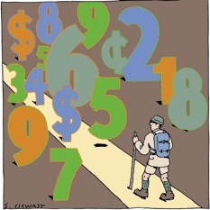 Elizabert Kolbert writes an interesting review in The New Yorker of several books on our irrational economic and political behaviors, the field of behavioral economics. It seems very likely that as politicians and governments become more knowledgeable about the patterns and emotional mechanisms governing our blunders, they will begin to nudge people towards more rational choices. The 'opt out' plans for increasing the numbers of people with health insurance plans or retirement savings are one example of this. Obama's campaign is making very good use of some basic neuro-economics and some of his people are aware of Westin's work (see my July 11 post), as well as Lakoff's (see Jan 31 post). Hillary doesn't seem to have a clue......
A test for Alzheimer's risk
 A news piece by Jennifer Couzin in the Feb. 22 Science notes that starting in about a month, for ~$400, you can send a saliva sample to Smart Genetics in Philadelphia for their "Alzheimer's Mirror" test that determines whether you have a variant of the APOE gene that indicates a risk of Alzheimer's that's 3 to 15 times higher than normal. The company plans plan to screen out those who seem emotionally unstable and provide a genetic counseling session by telephone before giving out APOE results.
Not surprisingly many physicians and researchers are expressing reservations about making this gene test widely available. What are the mental health consequences of being told you may get a disease that's neither preventable nor treatable and is invariably fatal? (It's the only genetic information that James Watson, the DNA discoverer who recently had his entire genome sequenced, kept secret.) Would it turn out that people who had this information were more likely experience depression?
An officer at Smart Genetics argues that knowing one is at higher risk might trigger practical responses, including regular memory screenings or making certain financial decisions such as buying long-term care insurance.
Monday, March 03, 2008
Chill out, and your wounds will heal faster.
 It is known that anger expression can be associated with increases in cortisol secretion and lowered immune function of the sort seen with other kinds of stress. Gouin et al. at Ohio State Univ. have examined the effect of anger on wound healing by following 98 community volunteers who agreed to receive a standardized blister wound on their non-dominant forearm. They found that wounds of those who expressed little anger or displayed anger in a controlled fashion healed more rapidly than the hotheads. The hotheads exhibited higher cortisol reactivity during the blistering procedure. This enhanced cortisol secretion was in turn related to longer time to heal.
 The data show more rapid wound healing in subjects with high anger control.
Measurement of the rate of transepidermal water loss (TEWL) through human skin provides a noninvasive method to monitor changes in the stratum corneum barrier function of the skin. TEWL was measured using a vapor pressure gradient estimation method. TEWL decreased as the barrier was restored; thus, monitoring of TEWL over time allowed objective evaluation of wound healing.
The Fires of Aging
Donna Holmes offers an interesting review with the title of this post, of Caleb Finch's new book "The Biology of Human Longevity." Here are a few edited clips from that review:
Metabolically speaking, we're all on fire. Current thinking in the biology of aging suggests that the normal processes cells use to burn fuel, providing energy for life, indirectly lead to much of the disease and disability that characterize aging in humans and other animals. Chemically unstable by-products of cellular oxidation--especially free oxygen radicals--can initiate the deterioration of cell membranes and macromolecules. As small "hits" causing cellular injury accumulate, the results can range from uncorrected mutations and cancers to forms of tissue damage leading to vascular pathology and Alzheimer's disease.
Oxidative damage remains a central player in the drama Fitch unfolds, but now it shares the stage with several lesser-known, equally important accomplices: inflammation, damage during development, and the hazards of overnutrition.Finch proposes that increases in brain size and the human life span over the past million years occurred in concert with changing nutritional priorities, slower developmental rates, and a tolerance for inflammation in "dirty, invasive, and stingy" prehistoric environments. The integration of more meat into the human diet, he argues, provided protein needed for larger brains but involved new physiological and genetic trade-offs between fitness and liability for long-term damage. This scenario provides a satisfying rationale for why variants of some genes for metabolizing animal fat that are linked to a human predilection for atherosclerosis, some cancers, and the amyloid plaques characteristic of Alzheimer's disease (such as those of the ApoE gene family) are not shared by our closest primate relatives.

Subscribe to:
Posts (Atom)