Exercise has many health benefits, including antidepressant actions in depressed human subjects, but the mechanisms underlying these effects have not been elucidated. We used a custom microarray to identify a previously undescribed profile of exercise-regulated genes in the mouse hippocampus, a brain region implicated in mood and antidepressant response. Pathway analysis of the regulated genes shows that exercise upregulates a neurotrophic factor signaling cascade that has been implicated in the actions of antidepressants. One of the most highly regulated target genes of exercise and of the growth factor pathway is the gene encoding the VGF nerve growth factor, a peptide precursor previously shown to influence synaptic plasticity and metabolism. We show that administration of a synthetic VGF-derived peptide produces a robust antidepressant response in mice and, conversely, that mutation of VGF in mice produces the opposite effects. The results suggest a new role for VGF and identify VGF signaling as a potential therapeutic target for antidepressant drug development.
Showing posts with label brain plasticity. Show all posts
Showing posts with label brain plasticity. Show all posts
Tuesday, January 22, 2008
Antidepressant effects of exercise - a mechanism
Here is an interesting bit from Hunsberger et al. in Nature Medicine, which suggests that a nerve growth factor pathway might be a target for antidepressant drug development (exercise might do the same thing, but depressed people usually aren't that keen on working out):

Friday, January 18, 2008
Exercise effects on brain and cognition
Hillman et al., provide an interesting review article (PDF here) that examines the positive effects of aerobic physical activity on cognition and brain function, at the molecular, cellular, systems and behavioral levels.
The results of a meta-analysis of the effects of fitness training on cognition showed that the benefits of fitness training on four different cognitive tasks were significant. As illustrated in the figure, fitness training has both broad and specific effects. The effects are broad in the sense that individuals in aerobic fitness training groups (represented by the red bars) showed larger fitness training effects across the different categories of cognitive processes illustrated on the x-axis. They are specific in the sense that fitness training effects were larger for some cognitive processes, in particular executive control processes, than for other cognitive processes.
Physical activity has been found to enhance cognition, with a selectively larger effect on executive control functions compared with other cognitive processes. Accordingly, brain structures that mediate executive functions would be expected to show disproportionate changes as a result of participation in physical activity. One such structure is the anterior cingulate cortex (ACC), which is part of the brain's limbic system and has connections with multiple brain structures that process sensory, motor, emotional and cognitive information. Two convergent lines of research indicate that physical activity exerts a substantial influence on the ACC and the concomitant executive processes that it mediates.


Thursday, January 17, 2008
Permanent Reincarnation
Here are some clips from an interesting essay by science writer Tor Norretranders:
My body is not like a typical material object, a stable thing. It is more like a flame, a river or an eddie. Matter is flowing through it all the time. The constituents are being replaced over and over again...98 percent of the atoms in the body are replaced every year. 98 percent! Water molecules stays in your body for two weeks (and for an even shorter time in a hot climate), the atoms in your bones stays there for a few months. Some atoms stay for years. But almost not one single atom stay with you in your body from cradle to grave...What is constant in you is not material. An average person takes in 1.5 ton of matter every year as food, drinks and oxygen. All this matter has to learn to be you. Every year. New atoms will have to learn to remember your childhood.
These numbers has been known for half a century or more, mostly from studies of radioactive isotopes. Physicist Richard Feynman said in 1955: "Last week's potatoes! They now can remember what was going on in your mind a year ago."
But digital media now makes it possible to think of all this in a simple way. The music I danced to as a teenager has been moved from vinyl-LPs to magnetic audio tapes to CDs to Pods and whatnot. The physical representation can change and is not important — as long as it is there. The music can jump from medium to medium, but it is lost if it does not have a representation. This physics of information was sorted out by Rolf Landauer in the 1960'ies. Likewise, out memories can move from potato-atoms to burger-atoms to banana-atoms. But the moment they are on their own, they are lost.We reincarnate ourselves all the time. We constantly give our personality new flesh. I keep my mental life alive by making it jump from atom to atom. A constant flow. Never the same atoms, always the same river. No flow, no river. No flow, no me...This is what I call permanent reincarnation: Software replacing its hardware all the time. Atoms replacing atoms all the time. Life. This is very different from religious reincarnation with souls jumping from body to body (and souls sitting out there waiting for a body to take home in).

Monday, January 14, 2008
Face perception after no experience of faces
This work really nails down the fact that face processing is a special perceptual process and is organized as such at birth, as contrasted with having its origin in a more general-purpose perceptual system that becomes specialized after frequent visual experiences. Sugita has studied face perception in monkeys reared with no exposure to faces. Here is his abstract, and one figure from the paper:
Infant monkeys were reared with no exposure to any faces for 6–24 months. Before being allowed to see a face, the monkeys showed a preference for human and monkey faces in photographs, and they discriminated human faces as well as monkey faces. After the deprivation period, the monkeys were exposed first to either human or monkey faces for a month. Soon after, the monkeys selectively discriminated the exposed species of face and showed a marked difficulty in regaining the ability to discriminate the other nonexposed species of face. These results indicate the existence of an experience-independent ability for face processing as well as an apparent sensitive period during which a broad but flexible face prototype develops into a concrete one for efficient processing of familiar faces.Figure: An infant monkey and her living circumstance. An infant monkey and a caregiver with (A) and without (B) a facemask. Both photos were taken after the face-deprivation period. (C) Toys placed in the monkey's home cage. (D) Decorations provided around the home cage.

Thursday, January 10, 2008
Compensatory neural plasticity in aging human brains.
Recent imaging studies have shown that seniors exhibit stronger brain activation than younger controls during the execution of various motor tasks. Old subjects activate the same regions as their younger counterparts, but to a larger extent, and they also activate additional regions that are not observed in the young subjects.
Heuninckx et al. examine the underlying neural mechanisms of this "overactivation" by determining whether it reflects compensation for various neural/behavioral deficits (e.g., neurodegeneration, attentional problems, reduction in sensory function, etc.) or whether it is due to de-differentiation (a generalized nonfunctional spread of activity attributable to deficits in neurotransmission, which in turn causes a decrease in the signal-to-noise ratio in neural firing and a loss of neural specialization). They compared brain activity in 24 older adults and 11 young controls during the performance of rhythmical hand–foot coordination tasks, whereby both limbs moved either in the same (iso-directional) or in the opposite (non-isodirectional, NONISODIR in the figure below) direction. Previous behavioral work had shown convincingly that the non-isodirectional pattern is more difficult and is produced with lower accuracy and stability than the iso-directional pattern. Activation in dedicated brain regions was correlated with motor performance in the elderly. According to the compensation hypothesis, the underlying rationale was that the over-activation would be larger in good than in poor motor performers, with the effect being more pronounced in more (non-isodirectional) than less (iso-directional) demanding coordination tasks. Conversely, the de-differentiation hypothesis assumed overactivation to be larger in poor than in successful motor performers because of nonfunctional neural irradiation. Thus, positive correlations between brain activation and motor performance were considered to reflect compensation, and negative correlations were considered to reflect de-differentiation.
They found that that coordination resulted in activation of classical motor coordination regions and also higher-level sensorimotor and frontal regions in the elderly. A positive correlation between activation level in these latter regions and motor performance was observed. This performance enhancing additional recruitment is consistent with the compensation hypothesis and reflects neuroplasticity at the systems level in the aging brain.

Figure: (Click to enlarge). Statistical parametric maps representing significantly larger activation in the old compared with the young group during the NONISODIR coordination mode, resulting from the following contrast: (NONISODIR – rest)old versus (NONISODIR – rest)young. L, Left hemisphere; R, right hemisphere. White arrows indicate brain regions that exhibit a significant correlation between brain activity level and coordination performance, as identified by a whole-brain multiple regression analysis. The graphics display each subject's BOLD response with respect to the within-cluster peak activation as a function of the inverse of the phase error (1/AE), with the younger subjects in blue and the older subjects in red.
Friday, December 28, 2007
Cognitive Recovery in Socially Deprived Young Children
With elaborate consideration of the ethical issues involved (commented on by Millum and Emanuel), Nelson et al. have compared the cognitive development of abandoned children reared in institutions to abandoned children placed in institutions but then moved to foster care (The Bucharest Early Intervention Project):
In a randomized controlled trial, we compared abandoned children reared in institutions to abandoned children placed in institutions but then moved to foster care. Young children living in institutions were randomly assigned to continued institutional care or to placement in foster care, and their cognitive development was tracked through 54 months of age. The cognitive outcome of children who remained in the institution was markedly below that of never-institutionalized children and children taken out of the institution and placed into foster care. The improved cognitive outcomes we observed at 42 and 54 months were most marked for the youngest children placed in foster care. These results point to the negative sequelae of early institutionalization, suggest a possible sensitive period in cognitive development, and underscore the advantages of family placements for young abandoned children.

Thursday, December 20, 2007
Race and IQ - a few crisp facts
 The debate over race and IQ seems endless and mind-numbing, usually generating more heat than light. A recent Op-Ed piece by Richard Nisbett, brief and to the point, collects several facts:
About 25 percent of the genes in the American black population are European, meaning that the genes of any individual can range from 100 percent African to mostly European. If European intelligence genes are superior, then blacks who have relatively more European genes ought to have higher I.Q.’s than those who have more African genes. But it turns out that skin color and “negroidness” of features — both measures of the degree of a black person’s European ancestry — are only weakly associated with I.Q. (even though we might well expect a moderately high association due to the social advantages of such features).
During World War II, both black and white American soldiers fathered children with German women. Thus some of these children had 100 percent European heritage and some had substantial African heritage. Tested in later childhood, the German children of the white fathers were found to have an average I.Q. of 97, and those of the black fathers had an average of 96.5, a trivial difference.
If European genes conferred an advantage, we would expect that the smartest blacks would have substantial European heritage. But when a group of investigators sought out the very brightest black children in the Chicago school system and asked them about the race of their parents and grandparents, these children were found to have no greater degree of European ancestry than blacks in the population at large.
.. a superior adoption study...looked at black and mixed-race children adopted by middle-class families, either black or white, and found no difference in I.Q. between the black and mixed-race children....children adopted by white families had I.Q.’s 13 points higher than those of children adopted by black families. The environments that even middle-class black children grow up in are not as favorable for the development of I.Q. as those of middle-class whites.
James Flynn, a philosopher and I.Q. researcher in New Zealand, has established that in the Western world as a whole, I.Q. increased markedly from 1947 to 2002. In the United States alone, it went up by 18 points. Our genes could not have changed enough over such a brief period to account for the shift; it must have been the result of powerful social factors. And if such factors could produce changes over time for the population as a whole, they could also produce big differences between subpopulations at any given time.
...interventions at every age from infancy to college can reduce racial gaps in both I.Q. and academic achievement, sometimes by substantial amounts in surprisingly little time. This mutability is further evidence that the I.Q. difference has environmental, not genetic, causes.
Video of independent leg movement controllers
Here, as a companion to my Sept. 20 post "Walking the walk" is a video illustrating the independent controllers of our right and left legs during walking.
Wednesday, December 12, 2007
Using Neuroimaging to Resolve the Psi Debate
Moulton and Kosslyn offer a study in J. Cog. Neurosci attempting to find evidence for the psi effect in brain imaging experiments. Here is their abstract, followed by some edited clips and a figure from the paper:
Parapsychology is the scientific investigation of apparently paranormal mental phenomena (such as telepathy, i.e., "mind reading"), also known as psi. Despite widespread public belief in such phenomena and over 75 years of experimentation, there is no compelling evidence that psi exists. In the present study, functional magnetic resonance imaging (fMRI) was used in an effort to document the existence of psi. If psi exists, it occurs in the brain, and hence, assessing the brain directly should be more sensitive than using indirect behavioral methods (as have been used previously). To increase sensitivity, this experiment was designed to produce positive results if telepathy, clairvoyance (i.e., direct sensing of remote events), or precognition (i.e., knowing future events) exist. Moreover, the study included biologically or emotionally related participants (e.g., twins) and emotional stimuli in an effort to maximize experimental conditions that are purportedly conducive to psi. In spite of these characteristics of the study, psi stimuli and non-psi stimuli evoked indistinguishable neuronal responses—although differences in stimulus arousal values of the same stimuli had the expected effects on patterns of brain activation. These findings are the strongest evidence yet obtained against the existence of paranormal mental phenomena.
In our experiment, participants played one of two roles: "sender" and "receiver." On each trial, sender participants viewed a randomly selected target stimulus from outside the scanner (see Figure), and tried to send this information to the receiver participant by mental means alone. While the senders were doing this, receiver participants completed a simple binary guessing task, and functional magnetic resonance imaging (fMRI) was used to monitor their brain activity. On each trial of the guessing task, the receivers sequentially viewed two stimuli, guessed which one was the stimulus being "sent" (i.e., the psi stimulus), and then saw the psi stimulus a second time. This paradigm allowed us simultaneously to test all three hypothesized mechanisms of psi: telepathy (i.e., "mind reading"), clairvoyance (i.e., direct sensing of remote events), and precognition (i.e., knowing future events). The sender served as the potential telepathic source, the sender's computer monitor served as the potential clairvoyance source, and the second presentation of the psi stimulus served as the potential precognition source.Figure 1 A schematic of one trial. In this trial for the receiver, the non-psi stimulus appears first and the psi stimulus second. The third stimulus presentation (feedback) in each trial is always the same as the psi stimulus. The sender sees only the psi stimulus for each trial.
The results support the null hypothesis that psi does not exist. The brains of our participants—as a group and individually—reacted to psi and non-psi stimuli in a statistically indistinguishable manner. Given the relatively large number of participants, the use of fixed-effects statistics, the extensive activation elicited separately by both types of stimuli, the subtle psychological effects revealed in the much smaller data set from a single participant, and the non-psi effects we documented on a group level using identical statistical criteria, a lack of statistical power does not reasonably explain our results. Even if the psi effect were very transient, as are many mental events, it should have left a footprint that could be detected by fMRI—as did the other subtle effects we detected. In particular, the large and massively significant activation revealed by our arousal contrast shows that that the psi effect, if it exists, must be substantially smaller than the effect of arousal on brain activity.
But what of the truism that one cannot affirm the null hypothesis? We note that some null results should be taken more seriously than others. ....Consider the possibility of water on Mars. If a set of close-up images of its surface failed to capture frozen lakes, few would accept the nonexistence of Martian water. Yet if a planetwide analysis of its subsurface soil content failed to show telltale signs of water, most would accept the null hypothesis of a Martian desert. Past null results from parapsychology are comparable to scattered snapshots of the surface in that they measure a small sample of outwardly observable variables. The current neuroimaging approach, however, seeks anomalous knowledge at its source, inside the brain, using methods validated by cognitive neuroscience. It is also exhaustive...the study incorporated methodological variables (e.g., biological and emotional relatedness of participants, evocative stimuli) widely considered to facilitate psi by parapsychologists. As such, the current null results do not simply fail to support the psi hypothesis: They offer strong evidence against it. If these results are replicated over a range of participants and situational contexts, the case will become increasingly strong, with as much certainty as is allowed in science, that psi does not exist.

Friday, November 16, 2007
The instinct to swarm
 Groups of social animals whose individual members follow simple sets of rules do surprising things. This NY Times article by Carl Zimmer in the Nov. 13 science section quotes Ian Couzin, a mathematical biologist at Princeton: “No matter how much you look at an individual army ant...you will never get a sense that when you put 1.5 million of them together, they form these bridges and columns. You just cannot know that.” The article notes the simple models that predict swarming behavior by setting the population density that which individuals switch from going their own way to following others. It also describes experiments using human subjects to test Couzin's models.
Many take our brains to be a more massive and complex version of the "hive minds" displayed by groups of bees, ants, birds and fish. Brain modelers assign relatively simple properties to their model neurons and then watch amazing patterns emerge when their whole society of neurons is fired up to interact.
Thursday, November 15, 2007
Exercise on the Brain
 Aamodt and Wang contribute an Op-Ed piece with the title of this post in the Nov. 8 New York Times. Their main message is that all of the 'brain exercise' programs that are marketed to counter the cognitive decline associated with aging are more expensive, complicated, and vastly less effective than vigorous daily exercise (not to suggest that these are competing alternatives, it is certainly best to do both). They note that while activities like solving puzzles or remembering lists can induce lasting changes in these specialized areas, physical exercise improves “executive function,” the set of abilities that allows you to select behavior that’s appropriate to the situation, inhibit inappropriate behavior and focus on the job at hand in spite of distractions. Executive function includes basic functions like processing speed, response speed and working memory, the type used to remember a house number while walking from the car to a party. They also note studies showing the numerous theraputic effects of exercise, such as delaying both the onset of dementia and the shrinking of the frontal cortex that occurs with age.
Wednesday, November 14, 2007
Society for Neuroscience meeting: news from the front lines
You might like to check out the Society for Neuroscience website, which offers very accessible information for general public, press, and educators. The site contains links to these topics from the recent annual meeting:
* Antidepressant Drugs, Exercise, Young Age, Even Food Intake, Frequency, and Type, Affect Generation of New Brain Cells
* Research Sheds Light on Brain Differences in Adolescents, Understanding their Impulsive, Risk-Taking Behavior
* Training, Sensory Substitution, Thought-Reading Computers, Sleep, and Molecular Imaging Advance Stroke Research
* Thoughts, Not Arms and Hands, Can Operate Machines: New Devices May Soon Improve Lives or Physically Handicapped
* New Research Explores Dietary Effects on Amyloid in Search for Ways To Prevent, Treat Alzheimer's Disease
* New Studies Find Potential Biomarker for PTSD, Make Gains in Understanding Disorder and Why it is Difficult To Treat
Thursday, November 08, 2007
Decision, Decisions
A recent Science Magazine issue has a special section on the underlying processes of decision-making. Here is Peter Stern's introduction to the section:
Who hasn't agonized over a major decision in life, whether to accept a job offer, move house, or perhaps switch research fields? We are confronted with a multitude of decisions on a daily basis. Many decisions are trivial and can be dealt with in seconds. However, others may have wider ramifications and can be excruciatingly complicated. In the past few years, our understanding of the underlying processes of decision-making has progressed markedly. This neuroscience special issue highlights some of the most exciting developments in this area.
Koechlin and Hyafil review recent experimental studies that provide new insights into the function and connectivity of the anterior prefrontal cortex, which forms the apex of the executive system underlying decision-making. The authors propose an original model of the anterior prefrontal function and provide a theoretical framework for addressing major unresolved issues and guiding future research on decision-making and higher cognition.
Human beings are highly social animals. Many of our decisions make sense only within a social environment. Sanfey outlines the advantages that can be gained by combining tasks and formal mathematical models from game theory with modern neuroimaging methods to characterize the processes that underlie social decision-making. He also summarizes recent research that offers good examples of how this neuroeconomic approach has already begun to illuminate our knowledge of this process.
Sometimes things can also go wrong in this complicated and well-balanced interplay between several brain regions. Paulus proposes that decision-making in psychiatric populations cannot be viewed simply as an alteration of the preference structure or the way individuals experience the outcome of the decision. Instead, it must be understood from the homeostatic balance perspective of the individual. Increased risk-taking behavior in drug addicts, for example, although maladaptive in the generic sense, may actually be adaptive for the substance user in a complex, highly unpredictable environment while attempting to respond to internal urges and cravings.
Decision theory has boomed in the past decade. Körding gives an overview of how decision theory, including normative/Bayesian approaches, can lead us to better understand the functions of the nervous system.

Wednesday, November 07, 2007
A Ramachandran lecture...
 In this excellent and engaging talk, Vilayanur Ramachandran discusses how brain damage can reveal the connection between internal structures of the brain and corresponding functions of the mind. Focus is on phantom limb pain, synesthesia (when people hear color or smell sounds), and the Capgras delusion (when brain-damaged people believe their closest friends and family have been replaced with imposters.)

Monday, November 05, 2007
Less SAD with more sun and serotonin
 Welberg offers a summary and review of work by Willeit et al. on the role of serotonin, and a serotonin transporter, in seasonal affective disorder. Here is an portion of the review:
Short, dark winter days put most of us in a gloomy mood, but in people with seasonal affective disorder (SAD), they can cause severe clinical depression. Fortunately, this depression can be treated with bright-light therapy (BLT), and it disappears altogether in summer. Willeit et al. now show that these changes in mood are associated with alterations in the efficiency of the serotonin (5-hydroxytryptamine) transporter (5-HTT) in the patients' blood platelets.
One theory of depression posits that impaired functioning of monoamine neurotransmitters, such as serotonin, causes the disorder, but it is unknown how this impairment might arise. Serotonin levels in the synapse are controlled by the 5-HTT, and Willeit and colleagues therefore investigated whether alterations in 5-HTT functioning might underlie depression in SAD.
The authors compared people with SAD with healthy volunteers, and assessed 5-HTT functioning in winter, after 4 weeks of BLT and in summer. They did this by measuring 5-HTT-mediated inward and outward transport in blood platelets (which are easily obtainable). In winter, both inward transport rate and outward transport were enhanced in the platelets of SAD patients compared with healthy controls. Importantly, these differences in platelet 5-HTT functioning disappeared after 4 weeks of BLT and were absent in summer. The number of 5-HTTs and their affinity for serotonin did not change with BLT or with the seasons, indicating that the increased 5-HTT inward transport that was found in SAD patients was due to increased efficiency of the transporter.
The authors also assessed the patients' depression levels at the three time points, using a structured interview. They found that post-treatment, both inward transport rate and outward transport correlated with depression scores in SAD patients. Moreover, patients whose depression did not decrease after treatment did not show a change in 5-HTT-mediated outward transport after treatment.
Friday, October 26, 2007
Brain changes after rehabilitation of congenital prosopagnosia
Another article on faces...Degutis et al. show MRI changes correlating with a recovery of enhanced amplitude of the N170 ERP (electroencephalogram event related potential)component in response to faces compared to objects after training of a subject with congenital prosopagnosia (face blindness).
We used functional magnetic resonance imaging (fMRI) and electroencephalography (EEG) to measure neural changes associated with training configural processing in congenital prosopagnosia, a condition in which face identification abilities are not properly developed in the absence of brain injury or visual problems. We designed a task that required discriminating faces by their spatial configuration and, after extensive training, prosopagnosic MZ significantly improved at face identification. Event-related potential results revealed that although the N170 was not selective for faces before training, its selectivity after training was normal. fMRI demonstrated increased functional connectivity between ventral occipital temporal face-selective regions (right occipital face area and right fusiform face area) that accompanied improvement in face recognition. Several other regions showed fMRI activity changes with training; the majority of these regions increased connectivity with face-selective regions. Together, the neural mechanisms associated with face recognition improvements involved strengthening early face-selective mechanisms and increased coordination between face-selective and nonselective regions, particularly in the right hemisphere.
Tuesday, October 09, 2007
Musicians have enhanced subcortical processing
Musical training is known to modify cortical organization. Musacchia et al. show that:
...such modifications extend to subcortical sensory structures and generalize to processing of speech. Musicians had earlier and larger brainstem responses than nonmusician controls to both speech and music stimuli presented in auditory and audiovisual conditions, evident as early as 10 ms after acoustic onset. Phase-locking to stimulus periodicity, which likely underlies perception of pitch, was enhanced in musicians and strongly correlated with length of musical practice. In addition, viewing videos of speech (lip-reading) and music (instrument being played) enhanced temporal and frequency encoding in the auditory brainstem, particularly in musicians. These findings demonstrate practice-related changes in the early sensory encoding of auditory and audiovisual information.
Monday, October 08, 2007
Plasticity and learning in the human mirror neuron system
I pass on a review by Welberg of an interesting study by Catmur et al. [Catmur, C., Walsh, V. & Heyes, C. Sensorimotor learning configures the human mirror system. Curr. Biol. 17, 1527–1531 (2007)]:
Neurons in the frontoparietal mirror system fire when one performs an action and when one observes someone else performing that same action. This system is thought to have a role in social cognition and, perhaps, in language acquisition. How the mirror neurons map sensory input onto its motor representation is unknown, but Catmur et al. demonstrate that these representations are not innate and can be altered by training.
The authors used transcranial magnetic stimulation (TMS) to stimulate the motor cortex of volunteers who were watching a video of a hand. When the volunteers watched the hand's index finger move, the TMS-induced motor-evoked potential (MEP) was greater in the abductor muscle of their own index finger than when they watched the little finger move; conversely, the MEP of their little finger's abductor muscle was greatest when they watched the little finger move. In other words, a muscle showed MEP enhancement when its owner watched a movement that is normally performed by that muscle; this 'mirror effect' is thought to reflect activity of the mirror neuron system.
Half of the volunteers then underwent incongruent training trials, in which they were asked to extend their little finger if the video showed a hand extending the index finger, and vice versa. People in congruent trials simply had to repeat the movement they saw in the video. The incongruent trials were assumed to train the mirror system to associate an observed finger movement with movement of a different finger of the volunteer's own hand.
Measuring TMS-induced MEPs after training, the authors found that volunteers who had undergone the incongruent training now showed greater MEPs in the muscle of one finger when watching the 'wrong' finger move in the video, indicating that a reversal of muscle-specific MEP enhancement during action observation had taken place.
This study shows that the 'mirror properties' of the mirror system are not innate. Rather, they can be trained, through sensorimotor experience, to transform observation into action. These findings imply that insufficient social interaction and consequent inadequate sensory experience might affect the development of the mirror neuron system, for example, in children with autism.
Thursday, September 20, 2007
Walking the Walk
A new study of human locomotion shows a pattern of changes in independent neural controllers for left and right legs. Here is the abstract from Choi and Bastian and a summary figure from the review by Miall.
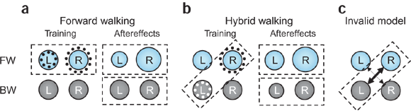
Human walking is remarkably adaptable on short and long timescales. We can immediately transition between directions and gait patterns, and we can adaptively learn accurate calibrations for different walking contexts. Here we studied the degree to which different motor patterns can adapt independently. We used a split-belt treadmill to adapt the right and left legs to different speeds and in different directions (forward versus backward). To our surprise, adults could easily walk with their legs moving in opposite directions. Analysis of aftereffects showed that walking adaptations are stored independently for each leg and do not transfer across directions. Thus, there are separate functional networks controlling forward and backward walking in humans, and the circuits controlling the right and left legs can be trained individually. Such training could provide a new therapeutic approach for correcting various walking asymmetries.Four neural systems are postulated, controlling forward (FW) and backward (BW) walking in left and right legs.
(a) In forward split-belt training, indicated by the dashed box, the right belt is faster than the left, inducing relative changes in the left and right forward-walking circuits (dotted circles). When walking on the tied-belt was tested after adaptation, an aftereffect was seen in forward walking, but not in backward walking. (b,c) In hybrid adaptive walking (b, diagonal dashed box), the left leg is on the slow backward belt and the right leg on the fast forward belt. This induced changes that were evident as aftereffects in both forward and backward walking, and that were compatible with this model of four functionally separate controllers, but were incompatible with a model (c, arrows) in which functional connections between these controllers are modified by learning.
Subscribe to:
Posts (Atom)