Wednesday, October 31, 2007

A Consciousness Debate

Christof Koch and Susan Greenfield offer a written version of their Oxford University Debate in the summer of 2006 in the Oct. 2007 issue of Scientific American. While they make a point of contrasting their models of the neuronal correlates of consciousness, I think a blending of the two may most closely approach the real situation.
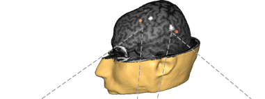
What happens in your brain when you see a dog, hear a voice, suddenly feel sad or have any other subjective experience?

KOCH'S MODEL
A coalition of pyramidal neurons linking the back and front of the cortex fires in a unique way. Different coalitions activate to represent different stimuli from the senses (left). In a mouse cortex (right) these pyramidal cells (green) lie in brain layer 5, surrounded by nonneuronal cells (blue).


GREENFIELD'S MODEL
Neurons across the brain fire in synchrony (green) and prevail until a second stimulus prompts a different assembly to arise (orange). Various assemblies coalesce and disband moment to moment, while incorporating feedback from the body. In a rat brain (bottom), an assembly in the cortex forms (a, b), peaks (c), then decays (d) within 0.35 second after the thalamus is electrically stimulated.

Christof Koch is professor of cognitive and behavioral biology at the California Institute of Technology, where he teaches and has conducted research on the neuronal basis of visual attention and consciousness for more than two decades.

Susan Greenfield is professor of pharmacology at the University of Oxford, director of the Royal Institution of Great Britain and member of the British Parliament's House of Lords. Her research focuses on novel brain mechanisms, including those underlying neurodegenerative diseases.

A new consiousness and philosophy of mind bibliography

David Chalmers and David Bourget are offering a more extensive online service, outlined in this message from ASSC (Assoc. for Sci.Stud. Cons.):
We are pleased to announce the launch of MindPapers, a new website
with a bibliography covering around 18000 published papers and online
papers in the philosophy of mind and the science of consciousness.
This site grew out of a combination of David Chalmers' bibliography in
philosophy of mind and his page of online papers on consciousness, but
it is much larger and has many new capacities, programmed by David
Bourget. The site address is:

http://consc.net/mindpapers/

There is also a separate front end for "Online Papers on
Consciousness". Where MindPapers now combines both offline published
papers and online papers from free and commercial sites, Online Papers
on Consciousness is devoted to free online papers (currently around
4700). It is based on the same database as MindPapers, but is
organized in a way to emphasize issues concerning consciousness and
cognitive science rather than the philosophy of mind. The address is

http://consc.net/online/

The MindPapers database contains 2773 papers on the philosophy of
consciousness (under 59 topics and subtopics) and 3917 papers on the
science of consciousness (under 71 topics and subtopics), as well as
thousands of papers on such related topics as perception,
intentionality, the philosophy of AI, and the philosophy of cognitive
science.

Capacities include (i) links and citation information throughout, (ii)
flexible navigation, display, and search options, (iii) the ability to
submit and edit entries, (iv) the capacity for automated off-campus
proxy access to commercial sites, and (v) a wealth of statistical
information.

We encourage everyone to try these sites to submit any relevant
material that we are missing (for a start, try searching on your own
name). There are tools on the site for submitting entries, as well as
for correcting entries and notifying us of any bugs and suggestions.

--David Chalmers and David Bourget
chalmers@anu.edu.au; david.bourget@anu.edu.au

Tuesday, October 30, 2007

Sleep deprivation diminishes recall of neutral and positive, but not of negative, events.

We remember emotional events, particularly negative ones, better than neutral events. Sterpenich et al. show that while consolidation of neutral and posititive memories is diminished by sleep deprivation, recall of negative events is less compromised. They show that after sleep deprivation, recollection of negative, potentially dangerous, memories recruits an alternate amygdalo-cortical network, which would keep track of emotional information despite sleep deprivation. Here is their description of the work:
Declarative memories, which can be consciously and verbally retrieved, are initially critically dependent on the hippocampus. However, reliable retrieval of long-term memory depends on a process of consolidation, which partly occurs during sleep, when memories are thought to be progressively transferred to long-term cortical stores. Because people tend to remember emotional memories better than neutral ones, we wondered whether the emotional significance of a memory would enhance its consolidation in a sleep-dependent manner. During a first session, participants viewed pictures with neutral and emotional content without realizing that their memory of the pictures and their content would be tested later (called incidental encoding). Three days later, during a functional MRI scanning session, subjects indicated whether they recognized previously viewed and new pictures. Half of the subjects were totally sleep deprived during the first post-encoding night, but all subjects slept as usual during the second and third post-encoding nights. We show here that the recollection of emotional stimuli elicited larger responses in the hippocampus and various cortical areas in the well-rested group than in the sleep-deprived group, suggesting that emotional significance boosts memory consolidation of the information during sleep. Interestingly, in sleep-deprived subjects, recollection of negative items recruited another network including the amygdala, as if an alternate consolidation process allowed them to keep track of negative, potentially dangerous, information despite the cognitive aftermath of sleep deprivation.

Biology and Health Inequality

PLoS Biology has ventured beyond its usual fare to publish several articles focusing on poverty, human development, and the environment. This article is from Eric Brunner. He points out several studies that demonstrate a direct psychosocial pathway to disease. It's precis: "Intriguing parallels between civil servant and nonhuman primate hierarchies suggest that highly stratified societies foster health inequalities. Determining how social differences translate into chronic disease remains a challenge, but neuroendocrine pathways appear to play a role."

Monday, October 29, 2007

Silent Minds

I want to point you to an excellent article by Jerome Groopman, with the title of this post, that appeared in a recent New Yorker Magazine. It describes recent work showing that brain imaging of some vegetative patients reveals responses to faces, and other visual and auditory inputs, that are indistinguishable from those of normal subjects (note: there are approximately 35,000 Americans in a vegetative state and another 280,000 in a minimally conscious state). Responses during various mental tasks, such as resolving ambiguous sentences or imagining playing a tennis game, can also be normal. This shows that an assumption held by doctors for decades - that vegetative patients lack capacity for conscious thought - is incorrect. Other vegetative patients (such as Terri Schiavo), in contrast, can show almost no cortical activity.

This all suggests a better medical definition of consciousness is required - such as the ability to report to ourselves or others the content of the representations in our brains, to sustain these representations over time and broadcast them broadly within the brain.

Evolution - with feeling....

A recent issue of American Scientist has a review by Robert Pennock of two books that attempt to show that a mechanistic Darwinian view of the world does not have to lead to a nihilistic ennui, but rather can satisfy our need to feel richness, purpose, and meaning.
With the familiar references to the "uncaring" Darwinian struggle, and the "mechanical" and "pitiless" action of natural selection, evolutionary biology has long been the obvious whipping boy for those who are uncomfortable with scientific naturalism. It is not just fundamentalist religious beliefs that motivate creationists' attacks on evolution; they are also driven by a deep existential angst—a fear that evolution renders the world pointless, emptying it of purpose, meaning and morality.
In "Darwin Loves You:Natural Selection and the Re-enchantment of the World" George Levine argues that evolution
if properly portrayed, is not only perfectly compatible with meaningfulness but provides a new basis for it...He makes the important point that at the same time that evolution pulls the rug out from under anthropocentrism (which is not only a smug but ultimately a dangerous attitude), it provides a foundation for a justifiable form of anthropomorphism. Darwin showed that humans are not the apex of creation but are one with the rest of the biological world, related to all living things through our common ancestors. This discovery allows us to find common ground with other animals without denigrating our humanness, Levine argues, permitting us to legitimately attribute human characteristics (albeit in simpler or incipient forms) to them. This provides an avenue to the re-enchantment of the world, for it shows we are not wrong to find in it a recognizably human notion of meaningfulness. It is wrong to see nature as cold and unfeeling; for those who understand evolutionary processes and relationships, the biological world becomes a warm and caring network of mutual interactions that are suffused with meaning. Levine is a romantic, but not a naive one; he does not close his eyes to those aspects of nature that are "red in tooth and claw," but shows how these need not negate the positive vision.
In "Evolution for Everyone: How Darwin's Theory Can Change the Way We Think about Our Lives" David Sloan Wilson, in the service of finding harmony between evolution and religion:
...discusses some of the evidence for his evolutionary hypothesis that religions are adaptive at the group level, providing practical benefits relating to the specific conditions the group is confronted with...Given the central importance of evolution in biology, the most extraordinary thing about the public's view, Wilson points out, is not that 50 percent don't believe it, but that nearly 100 percent haven't connected it to anything of importance in their lives. One of Wilson's chief goals—one he accomplishes admirably—is to demonstrate the relevance and value of evolutionary biology not just to scientists but to ordinary people. In story after engaging story, he conveys not only the sweep and the power of evolutionary thinking but the grandeur, as Darwin put it, of this view of life. By the end of the book, the reader understands Wilson's metaphor that evolution is an artist that has helped fashion the sculpture that is the living world.

Friday, October 26, 2007

The Outsourced Brain

I can't resist passing on this clever NY Times Op-Ed piece by David Brooks - on the subject of the dissolution of our individual intelligences into the mush of the infosphere.

Different takes on the social brain.

Gobbini et al. show that different types of mentalizing engage different brain regions. The abstract and a summary figure:
We compared two tasks that are widely used in research on mentalizing—false belief stories and animations of rigid geometric shapes that depict social interactions—to investigate whether the neural systems that mediate the representation of others' mental states are consistent across these tasks. Whereas false belief stories activated primarily the anterior paracingulate cortex (APC), the posterior cingulate cortex/precuneus (PCC/PC), and the temporo-parietal junction (TPJ)—components of the distributed neural system for theory of mind (ToM)—the social animations activated an extensive region along nearly the full extent of the superior temporal sulcus, including a locus in the posterior superior temporal sulcus (pSTS), as well as the frontal operculum and inferior parietal lobule (IPL)—components of the distributed neural system for action understanding—and the fusiform gyrus. These results suggest that the representation of covert mental states that may predict behavior and the representation of intentions that are implied by perceived actions involve distinct neural systems. These results show that the TPJ and the pSTS play dissociable roles in mentalizing and are parts of different distributed neural systems. Because the social animations do not depict articulated body movements, these results also highlight that the perception of the kinematics of actions is not necessary to activate the mirror neuron system, suggesting that this system plays a general role in the representation of intentions and goals of actions. Furthermore, these results suggest that the fusiform gyrus plays a general role in the representation of visual stimuli that signify agency, independent of visual form.

Figure - Locations of loci of activations associated with ToM, social animation, and biological motion tasks, projected onto the left and right lateral surfaces of the brain.

Brain changes after rehabilitation of congenital prosopagnosia

Another article on faces...Degutis et al. show MRI changes correlating with a recovery of enhanced amplitude of the N170 ERP (electroencephalogram event related potential)component in response to faces compared to objects after training of a subject with congenital prosopagnosia (face blindness).
We used functional magnetic resonance imaging (fMRI) and electroencephalography (EEG) to measure neural changes associated with training configural processing in congenital prosopagnosia, a condition in which face identification abilities are not properly developed in the absence of brain injury or visual problems. We designed a task that required discriminating faces by their spatial configuration and, after extensive training, prosopagnosic MZ significantly improved at face identification. Event-related potential results revealed that although the N170 was not selective for faces before training, its selectivity after training was normal. fMRI demonstrated increased functional connectivity between ventral occipital temporal face-selective regions (right occipital face area and right fusiform face area) that accompanied improvement in face recognition. Several other regions showed fMRI activity changes with training; the majority of these regions increased connectivity with face-selective regions. Together, the neural mechanisms associated with face recognition improvements involved strengthening early face-selective mechanisms and increased coordination between face-selective and nonselective regions, particularly in the right hemisphere.

Thursday, October 25, 2007

The male chill-out after sex: role for brain oxytocin

Here is an intriguing account from Waldherr and Neumann:
Sexual activity and mating are accompanied by a high level of arousal, whereas anecdotal and experimental evidence demonstrate that sedation and calmness are common phenomena in the postcoital period in humans. These remarkable behavioral consequences of sexual activity contribute to a general feeling of well being, but underlying neurobiological mechanisms are largely unknown. Here, we demonstrate that sexual activity and mating with a receptive female reduce the level of anxiety and increase risk-taking behavior in male rats for several hours. The neuropeptide oxytocin has been shown to exert multiple functions in male and female reproduction, and to play a key role in the regulation of emotionality after its peripheral and central release, respectively. In the present study, we reveal that oxytocin is released within the brain, specifically within the hypothalamic paraventricular nucleus, of male rats during mating with a receptive female. Furthermore, blockade of the activated brain oxytocin system by central administration of an oxytocin receptor antagonist immediately after mating prevents the anxiolytic effect of mating, while having no effect in nonmated males. These findings provide direct evidence for an essential role of an activated brain oxytocin system mediating the anxiolytic effect of mating in males.

It's in the Eyes!

Another curious bit on our brain's specialization for recognizing faces, noting the central role of the eyes. The abstract and a figure:
Unlike most other objects that are processed analytically, faces are processed configurally. This configural processing is reflected early in visual processing following face inversion and contrast reversal, as an increase in the N170 amplitude, a scalp-recorded event-related potential. Here, we show that these face-specific effects are mediated by the eye region. That is, they occurred only when the eyes were present, but not when eyes were removed from the face. The N170 recorded to inverted and negative faces likely reflects the processing of the eyes. We propose a neural model of face processing in which face- and eye-selective neurons situated in the superior temporal sulcus region of the human brain respond differently to the face configuration and to the eyes depending on the face context. This dynamic response modulation accounts for the N170 variations reported in the literature. The eyes may be central to what makes faces so special.

Figure - Simplified neural model of early face processing. Three sources are simultaneously active around 170 msec poststimulus onset. One source in the superior temporal sulcus (STS) region with a radial orientation generates the ERP N170 component. The combination of tangential sources in the fusiform gyrus (FG) and middle occipital gyrus (MOG) generates the MEG M170. The dynamic response modulation of eye- and face-selective neurons within the STS accounts for inversion and CR effects on the face N170 amplitude and for the other existing ERP data on the N170. The + signs represent the amount of activation of the neurons. The absence of + signs signifies that the neurons are not responding.

Wednesday, October 24, 2007

Our visual system is tuned to animals.

New et al. argue that the human attention system evolved category-specific selection criteria to monitor animals (including humans) in the environment. Ohman gives a nice commentary that puts the work in perspective (PDF here) Below is the abstract and a figure from New et.al. (PDF of article here):
Visual attention mechanisms are known to select information to process based on current goals, personal relevance, and lower-level features. Here we present evidence that human visual attention also includes a high-level category-specialized system that monitors animals in an ongoing manner. Exposed to alternations between complex natural scenes and duplicates with a single change (a change-detection paradigm), subjects are substantially faster and more accurate at detecting changes in animals relative to changes in all tested categories of inanimate objects, even vehicles, which they have been trained for years to monitor for sudden life-or-death changes in trajectory. This animate monitoring bias could not be accounted for by differences in lower-level visual characteristics, how interesting the target objects were, experience, or expertise, implicating mechanisms that evolved to direct attention differentially to objects by virtue of their membership in ancestrally important categories, regardless of their current utility.

Sample stimuli with targets circled. Although they are small (measured in pixels), peripheral, and blend into the background, the human (A) and elephant (E) were detected 100% of the time, and the hit rate for the tiny pigeon (B) was 91%. In contrast, average hit rates were 76% for the silo (C) and 67% for the high-contrast mug in the foreground (F), yet both are substantially larger in pixels than the elephant and pigeon. The simple comparison between the elephant and the minivan (D) is equally instructive. They occur in a similar visual background, yet changes to the high-contrast red minivan were detected only 72% of the time (compared with the smaller low-contrast elephant's 100% detection rate).


Brain location of verbal information storage varies between people

A group at Wisconsin has made the interesting observation that group averaged analyses of the sort frequently reported in brain imaging studies may give misleading results if the brain location of the process being studied varies from one individual to the next. Here is their abstract:
What are the precise brain regions supporting the short-term retention of verbal information? A previous functional magnetic resonance imaging (fMRI) study suggested that they may be topographically variable across individuals, occurring, in most, in regions posterior to prefrontal cortex (PFC), and that detection of these regions may be best suited to a single-subject (SS) approach to fMRI analysis. In contrast, other studies using spatially normalized group-averaged (SNGA) analyses have localized storage-related activity to PFC. To evaluate the necessity of the regions identified by these two methods, we applied repetitive transcranial magnetic stimulation (rTMS) to SS- and SNGA-identified regions throughout the retention period of a delayed letter-recognition task. Results indicated that rTMS targeting SS analysis-identified regions of left perisylvian and sensorimotor cortex impaired performance, whereas rTMS targeting the SNGA-identified region of left caudal PFC had no effect on performance. Our results support the view that the short-term retention of verbal information can be supported by regions associated with acoustic, lexical, phonological, and speech-based representation of information. They also suggest that the brain bases of some cognitive functions may be better detected by SS than by SNGA approaches to fMRI data analysis.

Figure: Example from subject 7 of SS and SNGA rTMS targets (orange markers; anterior, SNGA; posterior, SS). White blobs on the brain are load-sensitive regions identified by the SS analysis, which have been merged onto this subject's high-resolution T1-weighted anatomical scan, and are visible at this depth of scalp "peeling."

Reverie

I'm incredulous that over 38,000 people have viewed a recording of Debussy's Reverie that I put on YouTube, and 79 people have made comments (which led me to record a second version). There was also a video response that I thought I would pass on, another video titled "Reverie":

Tuesday, October 23, 2007

Origin of language from cortical motor systems - evidence from fMRI imaging

Meister and Iacoboni offer some interesting observations supporting the idea that language evolved as an exaptation from motor cortical systems. Here is some of their text and a central figure from the paper.
It has been suggested that cortical neural systems for language evolved from motor cortical systems, in particular from those fronto-parietal systems responding also to action observation. While previous studies have shown shared cortical systems for action – or action observation - and language, they did not address the question of whether linguistic processing of visual stimuli occurs only within a subset of fronto-parietal areas responding to action observation. If this is true, the hypothesis that language evolved from fronto-parietal systems matching action execution and action observation would be strongly reinforced

...functional magnetic resonance imaging (fMRI) was used while subjects watched video stimuli of hand-object-interactions and control photo stimuli of the objects and performed linguistic (conceptual and phonological), and perceptual tasks. Since stimuli were identical for linguistic and perceptual tasks, differential activations had to be related to task demands. The results revealed that the linguistic tasks activated left inferior frontal areas that were subsets of a large bilateral fronto-parietal network activated during action perception. Not a single cortical area demonstrated exclusive – or even simply higher - activation for the linguistic tasks compared to the action perception task.

The results show that linguistic tasks do not only share common neural representations but essentially activate a subset of the action observation network if identical stimuli are used. Our findings strongly support the evolutionary hypothesis that fronto-parietal systems matching action execution and observation were co-opted for language, a process known as exaptation.

Figure 3. a) cortical networks activated by the decision task relating to action observation vs rest (Vd-Perc vs rest, red) and action observation vs perceptual decisions on photos of the same objects (Vd-Perc vs Ph-Perc, blue). The large bihemispheric networks found for both contrasts were very similar, suggesting that the fMRI activations found here mainly were related to action observation and not to processes of decision making or object perception required during these tasks, as well. b) Cortical networks activated during the phonological (blue) and the conceptual decision task (red) on photos of manipulable objects. The networks activated by these two linguistic tasks were entirely part of the action observation network depicted in Fig. 3a, in accordance with the hypothesis that development of language out of the mirror neuron system was driven by a process of exaptation.

How potential reward biases our attention

Our eyes dart about (make saccades) rapidly as we view a visual scene - without these small rapid movements we are blind. Milsteen and Doris have made the fascinating observation that how rapidly we make these small movements can be influenced by the potential reward value of the visual target. Here is the work, in abstract-speak:
Basing higher-order decisions on expected value (reward probability x reward magnitude) maximizes an agent's accruement of reward over time. The goal of this study was to determine whether the advanced preparation of simple actions reflected the expected value of the potential outcomes. Human subjects were required to direct a saccadic eye movement to a visual target that was presented either to the left or right of a central fixation point on each trial. Expected value was manipulated by adjusting the probability of presenting each target and their associated magnitude of monetary reward across 15 blocks of trials. We found that saccadic reaction times (SRTs) were negatively correlated to the relative expected value of the targets. Occasionally, an irrelevant visual distractor was presented before the target to probe the spatial allocation of saccadic preparation. Distractor-directed errors (oculomotor captures) varied as a function of the relative expected value of, and the distance of distractors from, the potential valued targets. SRTs and oculomotor captures were better correlated to the relative expected value of actions than to reward probability, reward magnitude, or overall motivation. Together, our results suggest that the level and spatial distribution of competitive dynamic neural fields representing saccadic preparation reflect the relative expected value of the potential actions.

Monday, October 22, 2007

Language Evolution: An invisible hand.

A slightly edited "Editor's Summary" from the Oct. 11 Nature:
As a language evolves, grammatical rules emerge and exceptions die out. Lieberman et al. have calculated the rate at which a language grows more regular, based on 1,200 years of English usage. Of 177 irregular verbs, 79 became regular in the last millennium. And the trend follows a simple rule: a verb's half-life scales as the square root of its frequency. Irregular verbs that are 100 times as rare regularize 10 times faster. The emergence of a rule (such as adding –ed for the past tense) spells death for exceptional forms...In a separate study, Pagel et al. looked at changing word meanings. Across the Indo-European languages, words like 'tail' or 'bird' evolve rapidly and are expressed by many unrelated words. Others, like 'two', are expressed by closely related word forms across the whole language family. Data from over 80 modern languages show that the more a word is used, the less it changes.
And, from Fitch's review of the two papers:

The words of language are not inherited biologically, but are passed on culturally through learning. This process of 'cultural evolution' generates a hierarchical tree of relationships among languages, here illustrated by the Indo-European family. Just as descent with modification in biological evolution (phylogeny) leads to phylogenetic trees, so the analogous process in language change (glossogeny) can lead to glossogenetic trees.
Where should we look to gain a deeper understanding of the invisible hand in the cultural evolution of language? A promising future direction is provided by recent attempts to fuse theoretical models of cultural evolution to experimental investigations of social learning in the laboratory. Experimental investigations of 'iterated learning' — similar to the game of Chinese whispers, where one participant's output serves as input for the next — can provide empirical data to inspire, and constrain, our theories. Sophisticated new theoretical models enable language-learning 'agents' to have both innate biases (in the form of so-called bayesian priors) and powerful statistical learning systems capable of discovering and using environmental regularities. Such models demonstrate the possibility of a very indirect and sometimes non-intuitive relationship between the regularities emerging at the level of a whole population and the underlying generating forces. These forces are individual behaviour and learning (social usage) and innate constraints (in Chomsky's terms, a 'language acquisition device', often called universal grammar).

..some of the most persistent 'cultural replicators' — memes — evolve as slowly as some genes. By documenting and quantifying such effects, this work opens the door to a diverse range of theoretical and empirical investigations. If there is ever to be a science of memetics o rival that of genetics, it should proceed along these lines: combining careful quantitative analysis of well-documented linguistic changes with sophisticated theoretical models capable of taking into account the multilayered complexity of cultural evolution.

Emotional enhancement of memory and learning - a molecular mechanism.

Hu et al. have performed an interesting study of how the rush of noradrenaline during an emotional experience might enhance our ability to recall that experience. It causes a long term enhancement of the activity of nerve synapses using the neurotransmitter glutamate by stimulating the phosphorylation of a particular type of glutamate receptor. I'm afraid the molecular details will only make sense to those of you who know something about the chemistry of nerve transmission, but here they are, as outlined in the article's abstract:
Emotion enhances our ability to form vivid memories of even trivial events. Norepinephrine (NE), a neuromodulator released during emotional arousal, plays a central role in the emotional regulation of memory. However, the underlying molecular mechanism remains elusive. Toward this aim, we have examined the role of NE in contextual memory formation and in the synaptic delivery of GluR1-containing α-amino-3-hydroxy-5-methyl-4-isoxazoleproprionic acid (AMPA)-type glutamate receptors during long-term potentiation (LTP), a candidate synaptic mechanism for learning. We found that NE, as well as emotional stress, induces phosphorylation of GluR1 at sites critical for its synaptic delivery. Phosphorylation at these sites is necessary and sufficient to lower the threshold for GluR1 synaptic incorporation during LTP. In behavioral experiments, NE can lower the threshold for memory formation in wild-type mice but not in mice carrying mutations in the GluR1 phosphorylation sites. Our results indicate that NE-driven phosphorylation of GluR1 facilitates the synaptic delivery of GluR1-containing AMPARs, lowering the threshold for LTP, thereby providing a molecular mechanism for how emotion enhances learning and memory.

Saturday, October 20, 2007

A Wisconsin fall...

My partner and I went driving and walking in the Wisconsin countryside yesterday afternoon - here is an apple orchard, and the ice age trail.

The well-tempered web

I wanted to pass on this link to an engaging article by New Yorker music critic Alex Ross, on his experience with classical music culture on the web. The Internet may be killing the pop CD, but it's helping classical music. The article contains very useful links to many music sites.