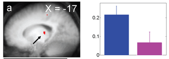
Figure - activity increase in left thalamus.
"We exposed 15 participants to short duration (50 s) monochromatic violet (430 nm), blue (473 nm), and green (527 nm) light exposures of equal photon flux (1013ph/cm2/s) while they were performing a working memory task in fMRI. At light onset, blue light, as compared to green light, increased activity in the left hippocampus, left thalamus, and right amygdala. During the task, blue light, as compared to violet light, increased activity in the left middle frontal gyrus, left thalamus and a bilateral area of the brainstem consistent with activation of the locus coeruleus.
These results support a prominent contribution of melanopsin-expressing retinal ganglion cells to brain responses to light within the very first seconds of an exposure. The results also demonstrate the implication of the brainstem in mediating these responses in humans and speak for a broad involvement of light in the regulation of brain function."
This blog reports new ideas and work on mind, brain, behavior, psychology, and politics - as well as random curious stuff. (Try the Dynamic Views at top of right column.)
Monday, December 10, 2007
Blue light changes our brains
A new light sensitive system has recently been discovered in the ganglion cells of the retinas, which send signals to the rest of the brain. These cells contain light sensitive melanopsin, most sensitive to blue wavelengths between 460 and 480 nm. The responses triggered by blue light takes seconds to develop and persist for minutes, unlike the rapid and transient responses of our rod and cone photoreceptor cells. Recent work has shown that brain activity related to a working memory task is maintained (or even increased) by blue (470 nm) monochromatic light exposure, whereas it decreases under green (550 nm) monochromatic light exposure. Vandewalle et al. now show that activation of this system causes changes in brain areas related to working memory:
Blog Categories:
attention/perception,
memory/learning
Subscribe to:
Post Comments (Atom)
No comments:
Post a Comment