Nakazawa et al. started by noting that increased expression of monocyte chemoattractant protein 1 (MCP-1) has been reported in vitreous humor samples of patients with RD (retinal detachment) and diabetic retinopathy as well as in the brain tissues of patients with neurodegenerative diseases, including Alzheimer's disease and multiple sclerosis. They moved on to do experiments showing that that MCP-1 plays a critical role in mediating photoreceptor apoptosis (cell death) in an experimental (mouse) model of RD. RD led to increased MCP-1 expression in the Müller glia and increased CD11b+ macrophage/microglia in the detached retina. An MCP-1 blocking antibody greatly reduced macrophage/microglia infiltration and RD-induced photoreceptor apoptosis. Confirming these results, MCP-1 gene-deficient mice showed significantly reduced macrophage/microglia infiltration after RD and very little photoreceptor apoptosis.
The work shows that MCP-1 expression and subsequent macrophage/microglia infiltration and activation are critical for RD-induced photoreceptor apoptosis. This pathway may be an important therapeutic target for preventing photoreceptor apoptosis in RD and other CNS diseases that share a common etiology.
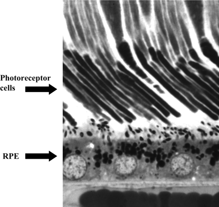
Photomicrograph showing the close physical relationship between the photoreceptor cells of the retina and the retinal pigment epithelium (RPE). There is constant metabolic exchange between the two cell systems.

Diagram of a cross-section of the eye with retinal detachment (open arrow) and retina hole (solid arrow). Fluid exchange between the subretinal space and vitreous cavity through the hole compromises metabolic exchange between the retina and retinal pigment epithelium.
No comments:
Post a Comment