We propose a new theory of infant pointing involving multiple layers of intentionality and shared intentionality. In the context of this theory, we argue and present evidence for a rich interpretation of prelinguistic communication, that is, one which posits that when 12-month-old infants point for an adult they are in some sense trying to influence her intentional/mental states. Moreover, we also argue and present evidence for a deeply social view in which infant pointing is best understood - on many levels and in many ways - as depending on uniquely human skills and motivations for co-operation and shared intentionality (e.g., joint intentions and attention with others). We conclude with a defense of the claim that children's initial skills of linguistic communication emerge on the heels of their initial pointing gestures because these two forms of interpersonal communication share a common social-cognitive, social-motivational infrastructure.
Tuesday, June 19, 2007
Human infant pointing, precursor to language?
From Tomasello's group, work with Malinda Carpenter and Ulf Liszkowski on "A New Look at Infant Pointing" (PDF here).
Music in Reuben's tube
Almost anything about music attracts my attention, and I thought this was a fascinating demonstration of standing waves and sound visualization.
The ‘when’ pathway of the right parietal lobe
Battelli et al provide a nice review (PDF here) in the May issue of Trends in Cognitive Sciences:
The order of events, whether two events are seen as simultaneous or successive, sets the stage for the moment-to-moment interpretation of the visual world. Evidence from patients who have lesions to the parietal lobes and transcranial magnetic stimulation studies in normal subjects suggest that the right inferior parietal lobe underlies this analysis of event timing. Judgment of temporal order, simultaneity and high-level motion are all compromised following right parietal lesions and degraded after transcranial magnetic stimulation over the right parietal but not elsewhere. The results suggest that the right parietal lobe serves as part of a when pathway for both visual fields. We propose that the disruption of this mechanism is the underlying cause of a wide range of seemingly unrelated tasks being impaired in right parietal patients.Figure. The when pathway. The when pathway is represented in the brain. This pathway is lateralized in the right hemisphere. Information from the primary visual cortex (V1) travels along the dorsal pathway (spatial perception, determining where objects are) or the ventral pathway (object recognition, determining what objects are), according to the classical subdivision that has been proposed based on animal models. A third pathway coming from V1 is dedicated to using time information to identify objects (e.g. determining when objects appeared or disappeared). Here, the temporoparietal junction (TPJ; considered the most common substrate of neglect) is identified as a core anatomical locus, within the inferior parietal lobe (IPL); however, the when pathway is likely to include a bigger network of areas, including the right angular gyrus (Ang), the supramarginal gyrus (Smg) and the posterior superior temporal sulcus (included in the superior temporal gyrus, STG). All these areas are often involved in the cortical lesion of right parietal patients. The intraparietal sulcus (IPS) separates the IPL from the superior parietal lobe (not labeled). The middle temporal area MT+ is reported in yellow (also called the motion area, highly specialized in detecting and discriminating moving stimuli).

Monday, June 18, 2007
Our brains putting the brakes on... "free won't" versus "free will"
One effort to rescue free will from the implication's of Libet's famous experiment (showing brain activity underlying an action starts before we are of aware of intending that action) has been to say "OK, even if "it" has started the action before "we" intended it, we can still shut it off. We still have "free won't."
Aron et al have now shown that the ability to stop motor reponses may involve a "hyperdirect" pathway between the right inferior frontal cortex and the basal ganglia (PDF of article here). Here is their abstract, followed by a figure from the paper:
Aron et al have now shown that the ability to stop motor reponses may involve a "hyperdirect" pathway between the right inferior frontal cortex and the basal ganglia (PDF of article here). Here is their abstract, followed by a figure from the paper:
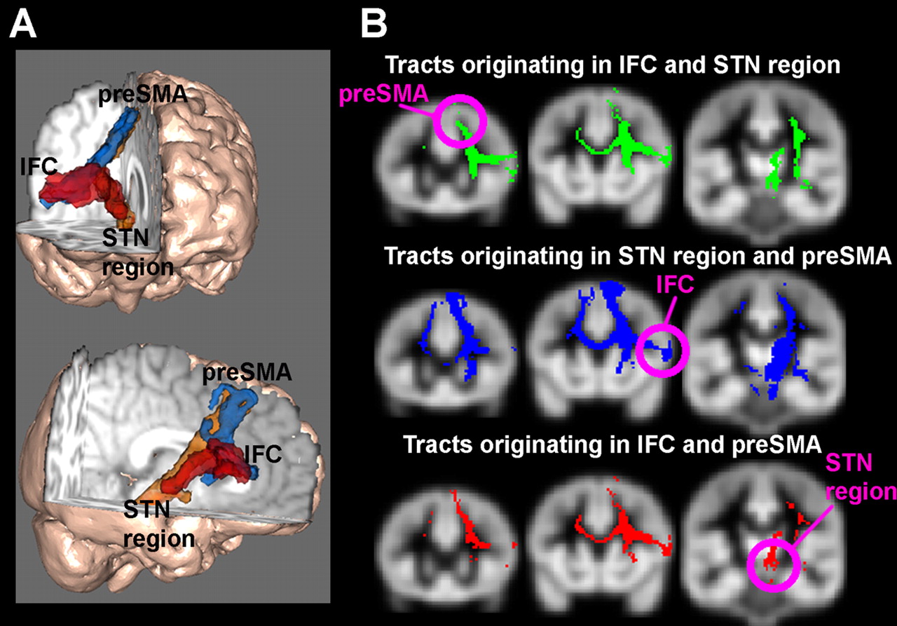
The ability to stop motor responses depends critically on the right inferior frontal cortex (IFC) and also engages a midbrain region consistent with the subthalamic nucleus (STN). Here we used diffusion-weighted imaging (DWI) tractography to show that the IFC and the STN region are connected via a white matter tract, which could underlie a "hyperdirect" pathway for basal ganglia control. Using a novel method of "triangulation" analysis of tractography data, we also found that both the IFC and the STN region are connected with the presupplementary motor area (preSMA). We hypothesized that the preSMA could play a conflict detection/resolution role within a network between the preSMA, the IFC, and the STN region. A second experiment tested this idea with functional magnetic resonance imaging (fMRI) using a conditional stop-signal paradigm, enabling examination of behavioral and neural signatures of conflict-induced slowing. The preSMA, IFC, and STN region were significantly activated the greater the conflict-induced slowing. Activation corresponded strongly with spatial foci predicted by the DWI tract analysis, as well as with foci activated by complete response inhibition. The results illustrate how tractography can reveal connections that are verifiable with fMRI. The results also demonstrate a three-way functional–anatomical network in the right hemisphere that could either brake or completely stop responses.Figure. Diffusion-weighted tractography results. A, 3-D rendering of the tracts between the right IFC, the right preSMA, and the right STN region. B, Triangulation method for determining the third point in a network from the other two. Tracts originating in one brain area are overlaid on tracts originating from another. The overlap is superimposed on a gray matter mask in standard space. Tracts clearly overlap in the white matter space, but the overlap in gray matter is fairly unique: the preSMA only for tracts originating in the IFC and STN regions; the IFC and anterior prefrontal cortex (not shown) for tracts originating in the preSMA and the STN region and the thalamus only for tracts originating in the preSMA and the IFC.
Laughing rats...
This from Panskepp. You might check out his other work on affiliative behavior and play. This article has relevant references and links.
Brain activation that reflects perceptual choices.
Thielscher and Pessoa (PDF here) have examined brain activation that reflects perceptual choices using fMRI during a two-choice perceptual discrimation task of fearful versus disgusted faces. From their abstract:
A review of this work by Tobler and Kalenscher (PDF here) provides a useful summary graphic:
 Figure 1. Top, Model of decision process. Bottom, Selected decision-related activations. Regions in orange predict whether a fearful or a disgusted face has been presented. Regions in blue predict whether the participant will choose "fearful" or "disgusted," and regions in yellow correlate with decision as operationalized by trial-to-trial changes in reaction time.
...Our analyses revealed that reporting a neutral face as "fearful" was associated with activation in a broad network of brain regions that process emotionally arousing stimuli, whereas reporting a neutral face as "disgusted" was associated with activation in a focused set of sites that included the putamen and anterior insula...fluctuations in fMRI amplitude for an individual participant could be used to reliably predict the perceptual choice of individual trials for that subject. In addition to the investigation of choice, we also isolated the neural correlates of decision making per se by using reaction time as an index of decision processes. Overall, our findings revealed that brain responses dynamically shifted according to perceptual choices. In addition, the neural correlates of decision making involved at least the anterior cingulate cortex, middle frontal gyrus, and inferior frontal gyrus/insula, consistent with recent proposals that decisions may emerge from distributed processes.
A review of this work by Tobler and Kalenscher (PDF here) provides a useful summary graphic:
 Figure 1. Top, Model of decision process. Bottom, Selected decision-related activations. Regions in orange predict whether a fearful or a disgusted face has been presented. Regions in blue predict whether the participant will choose "fearful" or "disgusted," and regions in yellow correlate with decision as operationalized by trial-to-trial changes in reaction time.
Friday, June 15, 2007
Searching MindBlog - keywords (labels) and search function now added
I have now cruised through the 500+ posts of MindBlog since it started up in Feb. of 2006 and done a cursory job of adding keywords to each. Using the labels list to the left, you can pull up all the posts in the areas of greatest interest to me. While I was tempted to distinguish many more categories, I thought this would get unwieldy and run the list too far down the screen, so I've limited the labels or keywords to about 35 items. Here is a list of labels I was also tempted to
add as I went through the old posts:
affiliative neuroscience
empathy
altruism
alzheimers
computers
modeling
amygdala
group selection
pain
human evolution
pheromones
brain development
placebo
gender
gay
anthropology
folk psychology
intelligence
addiction/drugs
depression
BUT, entering any of these in the blog search box provided by Google in the left column also gets you there. The Google search function proves to be very powerful for more focused searches, as for specific brain structures (insula, amygdala, whatever....)., or experiments using MRI imaging, etc.
Musical intervals in speech
Ross, Choi and Purves (PDF here) offer a fascinating study how vocal tract anatomy and vocal language sounds might explain why humans, across cultures, have created music using pitch intervals that divide octaves into the 12 tones of the chromatic scale:
Throughout history and across cultures, humans have created music using pitch intervals that divide octaves into the 12 tones of the chromatic scale. Why these specific intervals in music are preferred, however, is not known. In the present study, we analyzed a database of individually spoken English vowel phones to examine the hypothesis that musical intervals arise from the relationships of the formants in speech spectra that determine the perceptions of distinct vowels. Expressed as ratios, the frequency relationships of the first two formants in vowel phones represent all 12 intervals of the chromatic scale. Were the formants to fall outside the ranges found in the human voice, their relationships would generate either a less complete or a more dilute representation of these specific intervals. These results imply that human preference for the intervals of the chromatic scale arises from experience with the way speech formants modulate laryngeal harmonics to create different phonemes.The periodicity in speech sound stimuli is generated primarily by the repeating peaks of energy in the vocal air stream produced by oscillations of the vocal folds in the larynx. The intensity carried by the harmonic series produced in this way is altered, however, by the resonance frequencies of the rest of the vocal tract, which change dynamically in response to neurally controlled movements of the soft palate, tongue, lips and other articulators (see figure). These variable vocal tract resonances, called formants, modulate the harmonic series generated by the laryngeal oscillations by suppressing some harmonics more than others. When coupled with unvoiced speech sounds (consonants), this modulation by the formants creates the different voiced speech sounds that give rise to the semantic content in all human languages. With respect to vowel phones, only the first two formants have a major influence on the vowel perceived: artificially removing them from vowel phones makes vowel phonemes largely indistinguishable, whereas removing the higher formants has little effect on the perception of speech sounds. Indeed, the first and second formants of vowel sounds of all languages fall within well defined frequency ranges. The resonances of the first two formants are typically between approximately 200–1,000 Hz and approximately 800–3,000 Hz, respectively, their central values approximating the odd harmonics of the resonances of a tube approximately 17 cm in length open at one end, the usual physical model of the adult vocal tract in a relaxed state).
Figure - Ranges of the peak harmonic in the first two formants (F1 and F2) for eight American English vowels uttered as single words in an emotionally neutral manner. (A) Diagram of the human larynx and vocal tract; see Introduction for explanation. (B) Distribution of the peak harmonics selected as the index for the first and second formant for the five male participants. (C) Distribution for the five female participants. The somewhat smaller harmonic ranges for females are due to the higher average fundamental frequency of female speech. The mean fundamental frequency for male speakers was 109 Hz (SD = 10) and for female speakers 171 Hz (SD = 20).

When Lots of Money Makes You Feel Rich
I'm passing on a curious (and not too surprising) short piece by Alex Mindlin in the New York Times with the same title this post:


Anyone who has ever thrown around a stack of liras or rupees knows that people are sometimes more extravagant with currencies that have high face values. A paper recently published in The Journal of Consumer Research explored that effect...In one study, students in Hong Kong, when asked to allocate spending from an imaginary paycheck of 9,000 Hong Kong dollars, devoted an average of 532.35 of those dollars to food spending...Two weeks later, the students were asked to imagine that they had moved to the fictional country of Tristania, where a HongKong dollar equaled 18 Tristanian dollars, and therefore their pay was 162,000 Tristanian dollars. The students splurged, spending 30 percent more on food in real terms...The opposite effect was seen among students sent to an alternate Tristania where their paychecks were worth only 500 Tristanian dollars...“You feel more rich if you have more units of currency,” said Dilip Soman, a professor of marketing at the University of Toronto, and one of the paper’s authors...Earlier studies have been taken to suggest the opposite — that consumers are wary of spending in “numerous” foreign currencies, because they are put off by the high numbers on price tags. But Mr. Soman said that those studies had not taken budgeting into account.

Thursday, June 14, 2007
New cells in the brain require new experiences to live...
Leuner et al offer a nice review PDF here) of work by Tashiro et al. showing that survival of the thousands of new nerve cells that are born in the dentate gyrus of the hippocampus each day is enhanced if mice are exposed to new experiences during a critical window of time. Here is Tashiro et al.'s abstract, followed by a summary diagram from the Leuner review.
Neural circuits in the dentate gyrus are continuously modified by adult neurogenesis, whose level is affected by the animal's experience. However, it is not known whether this experience-dependent anatomical modification alters the functional properties of the dentate gyrus. Here, using the expression of immediate early gene products, c-fos and Zif268, as indicators of recently activated neurons, we show that previous exposure to an enriched environment increases the total number of new neurons and the number of new neurons responding to reexposure to the same environment. The increase in the density of activated new neurons occurred specifically in response to exposure to the same environment but not to a different experience. Furthermore, we found that these experience-specific modifications are affected exclusively by previous exposure around the second week after neuronal birth but not later than 3 weeks. Thus, the animal's experience within a critical period during an immature stage of new neurons determines the survival and population response of the new neurons and may affect later neural representation of the experience in the dentate gyrus. This experience-specific functional modification through adult neurogenesis could be a mechanism by which new neurons exert a long-term influence on the function of the dentate gyrus related to learning and memory.Here is a summary figure of the results (NeuN+ is a marker for nerve cells; BrdU is a marker for new neurons; Zif268 is a marker for increased neural activation.
Figure legend: Simplified schematic diagram of the results presented by Tashiro et al. (2007). Exposure to environmental enrichment during a critical period (1–3 weeks after BrdU administration; bottom) increased the survival of new neurons in the dentate gyrus (BrdU+/NeuN+). In contrast, new neuron survival was not enhanced in mice exposed to an enriched environment after the critical period (top). The authors also demonstrated that reexposure to the same experience of environmental enrichment at a later time enhanced neuronal activation (BrdU+/NeuN+/Zif268+; bottom). Increased neuronal activation did not occur when mice were only given the initial exposure or were reexposed to a different experience of water maze training.

More on the mirror "hearing-doing" system
Here is a more extended commentary on a paper I mentioned in my Jan. 30 post "The pianist in the mirror.." D'Ausilio (PDF here) discusses this work of Lahav et al. showing an auditory mirror area in the left inferior frontal gyrus for complex and newly acquired actions. In addition to rote auditory-motor mapping for learning and online execution control, this mirror mechanism might subserve other evolutionary critical functions like action recognition, and interindividual emotional resonance. This auditory mirror-like property seems indeed to be valid for a wide range of functions that in turn elicit very different behaviors.
Wednesday, June 13, 2007
Who watches the watcher?
 I recommend reading Adami's review (PDF here) of Douglas Hofstadter's recent book on consciousness "I am a Strange Loop," Basic Books, New York, 2007. A few excerpts:
Hofstadter's explanation of human consciousness is disarmingly simple...the main idea is simply that our feeling of a conscious "I" is but an illusion created by our neuronal circuitry: an illusion that is only apparent at the level of symbols and thoughts, in much the same way as the concepts of pressure and temperature are only apparent at the level of 10^23 molecules but not the level of single molecules. In other words, Hofstadter denies consciousness an element of ontological reality, without denying that our thoughts and feelings, pains and longings have an "inner reality" when we have them.
Hofstadter's book and Koch's recent "The Quest for Consciousness" make for an interesting juxtaposition. Each addresses the same problem but entirely on different levels. Yet both authors reach some of the same conclusions, sometimes using precisely the same metaphor (as when they compare the activity of "making up one's mind" in terms of a voting process). In the end, both authors could have profited from peeking at each other's arsenal: Hofstadter would probably be delighted to see some of the putative neural underpinnings of consciousness, to peer underneath the strange loop as it were, at the inordinately complex firework and the neuroanatomy that supports it. For his part, Koch would no doubt appreciate the computational trick that Gödel incompleteness plays on us, as well as the developmental aspect of consciousness that Hofstadter advocates.
The Accidental Mind
Geory Striedter writes a review of David Linden's new book "The Accidental Mind: How Brain Evolution Has Given Us Love, Memory, Dreams, and God" ..Harvard University Press: 2007. (PDF of the review is here) Some clips:
The human brain, and hence the human mind, is not an optimal, designed-from-scratch apparatus. Rather, it is an imperfect amalgam of shoddy components. That is the central thesis of David Linden's new book The Accidental Mind. Neurons are slow, leaky, and unreliable — hardly ideal computing elements. The whole brain, too, is not designed to the plan of some omnipotent engineer. Instead, evolution has endowed it with plenty of 'anachronistic junk'. Which is why, according to Linden, our minds often distort reality and can lead us to act foolishly. For example, when you reach out to touch something, your brain filters out what it expects. This selective neglect of expected input allows us to focus on unexpected stimuli, but it can be counterproductive. It may explain, for instance, why pushing and shoving confrontations tend to escalate. When someone pushes you, you feel it more than when you push the other with the same force, because the sensation caused by your own push is largely, though unconsciously, expected by your brain.
Linden tells his story well, in an engaging style, with plenty of erudition and a refreshing honesty about how much remains unknown. The book should easily hold the attention of readers with little background in biology and no prior knowledge of brains. It would make an excellent present for curious non-scientists and a good book for undergraduates who are just entering into the brain's magic menagerie. Even readers trained in neuroscience are likely to enjoy the many tidbits of rarely taught information — on love, sex, gender, sleep and dreams — that spice up Linden's main argument.

Tuesday, June 12, 2007
Our prefrontal control system is altered by unconscious stimuli.
Lau et al. (PDF here) used fMRI to test
..whether unconscious information can influence the cognitive control system in the human prefrontal cortex. Volunteers had to prepare to perform either a phonological judgment (whether the word is bisyllabic) or a semantic judgment (whether the word refers to concrete objects) on an upcoming word, based on the instruction given at the beginning of each trial. However, in some trials they were visually primed to prepare for the alternative (i.e., "wrong") task, and this impaired their performance. This priming effect is taken to depend on unconscious processes because the effect was present even when the volunteers could only discriminate the identity of the primes at chance level. Furthermore, the effect was stronger when the visibility of the prime was near zero than when the visibility of the prime was significantly higher. When volunteers were unconsciously primed to perform the alternative task, there was also decreased neural activity in the brain areas relevant to the instructed task and increased neural activity in the brain areas relevant to the alternative task, which shows that the volunteers were actually engaged in the wrong task, instead of simply being distracted. Activity in the mid-dorsolateral prefrontal cortex was also found to be associated with this unconscious priming effect. These results suggest that the cognitive control system in the prefrontal cortex is not exclusively driven by conscious information, as has been believed previously.Neural activity associated with priming. (Click to enlarge). Data were extracted from the brain areas that were previously found to be associated with the Phonological task (left ventral premotor area) and the Semantic task (left inferior frontal cortex and middle temporal gyrus). The location of these is schematically illustrated on the brain above. Here, task relevant means activity from the Semantic areas when the volunteers were instructed to perform the Semantic task, and activity from the Phonological areas when they were instructed to perform the Phonological task. Task irrelevant means the activity was extracted from the alternative areas, which was more important for the primed task than the instructed task. When the visibility of the primes was low (LoVis), which means the priming effect was strongest, activity in task-relevant areas was significantly reduced (p = 0.019), and activity in task-irrelevant areas was significantly increased (p = 0.045). This suggests that the volunteers were actually engaged in exercising the wrong neural circuits when they were primed to perform the wrong task. This effect is not present when the visibility of the primes was high (HiVis), suggesting that this effect could not be attributable to the degree of conscious perception of the prime. This is reflected by a three-way interaction between Task Relevance (i.e., activity in task relevant areas vs activity in task irrelevant areas), Congruency (Con; between prime and instruction), and Visibility (of the prime) (p = 0.040). InCon, Incongruency; n.s., not significant.
Mid-DLPFC and unconscious priming. (Click to enlarge) Lau et al. looked for activity in the brain that was associated with the unconscious priming effect in general, regardless of which task was explicitly cued, by testing for the interaction between Congruency (Con; between prime and instruction) and Visibility (of the prime). This test revealed activity in the mid-DLPFC (right; x = –38). This effect was specific to the Low-Visibility condition, in which volunteers did not consciously perceive the primes. InCon, Incongruency; n.s., not significant; HiVis, high visibility; LoVis, low visibility.


Monday, June 11, 2007
Use Viagra to recover from jet lag?
I always perk up when I see reports of new side effects of sildenafil (Viagra), because it is an inhibitor of one form of an enzyme, cyclic GMP phosphodiesterase, that my earlier research showed to be central to changing light into a nerve signal in the rod and cone cells of our retinas (check here if you are curious about this previous life..). One of the interesting side effects of viagra is on color vision. Here is another interesting bit from Agostino et al. in PNAS:
Mammalian circadian rhythms are generated by a master clock located in the suprachiasmatic nuclei and entrained by light-activated signaling pathways. In hamsters, the mechanism responsible for light-induced phase advances involves the activation of guanylyl cyclase, cGMP and its related kinase (PKG). It is not completely known whether interference with this pathway affects entrainment of the clock, including adaptation to changing light schedules. Here we report that cGMP-specific phosphodiesterase 5 is present in the hamster suprachiasmatic nuclei, and administration of the inhibitor sildenafil (3.5 mg/kg, i.p.) enhances circadian responses to light and decreases the amount of time necessary for reentrainment after phase advances of the light–dark cycle. These results suggest that sildenafil may be useful for treatment of circadian adaptation to environmental changes, including transmeridian eastbound flight schedules.
Subscribe to:
Posts (Atom)