Friday, November 30, 2007

Brain changes associated with congenital amusia

About four percent of the population has congenital amusia, a lifelong disability that prevents otherwise normal functioning individuals from developing basic musical skills. The condition has also been variously termed note deafness, tone deafness, or tune deafness. Here is an interesting abstract from Hyde et al. on the amusic brain, along with one figure from the paper:
Congenital amusia (or tone deafness) is a lifelong disorder characterized by impairments in the perception and production of music. A previous voxel-based morphometry (VBM) study revealed that amusic individuals had reduced white matter in the right inferior frontal gyrus (IFG) relative to musically intact controls. However, this VBM study also revealed associated increases in gray matter in the same right IFG region of amusics. The objective of the present study was to better understand this morphological brain anomaly by way of cortical thickness measures that provide a more specific measure of cortical morphology relative to VBM. We found that amusic subjects (n = 21) have thicker cortex in the right IFG and the right auditory cortex relative to musically intact controls (n = 26). These cortical thickness differences suggest the presence of cortical malformations in the amusic brain, such as abnormal neuronal migration, that may have compromised the normal development of a right frontotemporal pathway.

Figure - Group cortical thickness differences. Results from the statistical analysis of data from 21 amusics versus 26 controls are displayed at each vertex of the surface of a standardized brain in terms of a t statistical color map. A, Areas of significant thickness increases in the amusic brain relative to controls. B, areas of significant thickness decreases in the amusic brain relative to controls. Predicted group cortical thickness differences are in pink, and nonpredicted differences in green as follows: right superior precentral gyrus (a), right lateral occipital gyrus (b), right inferior precentral gyrus (c), right middle frontal gyrus (d), left inferior temporal gyrus (e), right anterior cingulate (f), right medial orbital frontal gyrus (g), right inferior temporal gyrus (h), left medial orbital frontal gyrus (i).

Roach Robots

Here is a moment of visual relief:

Thursday, November 29, 2007

Genetic variation in the enhancement of intelligence by breastfeeding.

It's the environment AND the genes...Caspi et al. provide a fascinating example of how genetic differences can moderate the effects of environmental influences on an individuals' health and behavior. The authors chose breastfeeding as the environmental exposure because the biological processes underlying its benefits for the developing brain are increasingly well understood. Here are some edited clips from the paper:
The predominant long-chain polyunsaturated fatty acids (LC-PUFAs) present in human milk, but not in cow's milk or most infant formulas, are docosahexaenoic acid (DHA; 22:6n-3) and arachidonic acid (AA or ARA; 20:4n-6). Substantial amounts of DHA and AA accumulate in the human brain during the first postnatal months, and infants who are breastfed have higher concentrations of DHA and AA than infants fed unsupplemented formulas. Evidence, in general, is consistent with the hypothesis that LC-PUFAs in breast milk may enhance cognitive development. In humans, children who are breastfed have higher IQs than children not fed breast milk, and this advantage persists into adulthood.

The authors chose to study people with two allelic variants of FADS2, which they termed C and G. FADS2 is an attractive candidate gene because of its role in the modification of dietary fatty acids. FADS2, located on chromosome 11q12.2, encodes the delta-6 desaturase that is the rate-limiting step on the metabolic pathway leading to AA and DHA production.

Examination of different cohorts of several thousand children from ongoing longitudinal studies in New Zealand, England and Wales revealed that breastfed children carrying the C allele showed a 6.4-IQ-point advantage relative to children not fed breast milk. GG homozygotes neither gained an advantage from breastfeeding nor suffered a disadvantage from not being fed breast milk. The authors ruled out alternative explanations of the finding involving gene–exposure correlation, intrauterine growth, social class, and maternal cognitive ability, as well as maternal genotype effects on breastfeeding and breast milk.

...the finding has implications for the public understanding of genetics. The pendulum of opinion surrounding nature versus nurture has swung back and forth, yielding global estimates of heritability versus "environmentality" that have overlooked the contribution of interactions between specific genes and specific experiences. To date, research on gene–environment interactions has been dominated by the search for genetic variants that increase disease susceptibility to environmental pathogens; for example, carriers of "short" 5-HTT alleles who encounter stressful life events are at risk of becoming depressed; carriers of "rapid" NAT2 alleles who eat red meat are at risk of developing colorectal cancer. However, genes are not only implicated in disease; here we have shown that a genetic variant (in FADS2) may also enhance a favorable response (increased IQ) to a salubrious exposure present throughout human ancestry (breastfeeding).

Pay me for my content!

This is the title of a brief Op-Ed piece by Jaron Lanier, a well known guru of the information age, in the Nov. 20 N.Y. Times. It is worth reproducing in full:
INTERNET idealists like me have long had an easy answer for creative types — like the striking screenwriters in Hollywood — who feel threatened by the unremunerative nature of our new Eden: stop whining and figure out how to join the party!..That’s the line I spouted when I was part of the birthing celebrations for the Web. I even wrote a manifesto titled “Piracy Is Your Friend.” But I was wrong. We were all wrong.

Like so many in Silicon Valley in the 1990s, I thought the Web would increase business opportunities for writers and artists. Instead they have decreased. Most of the big names in the industry — Google, Facebook, MySpace and increasingly even Apple and Microsoft — are now in the business of assembling content from unpaid Internet users to sell advertising to other Internet users. (Disclosure: I’m the scholar at large for Microsoft Live Labs, and I once was part of a company that Google bought.)

There’s an almost religious belief in the Valley that charging for content is bad. The only business plan in sight is ever more advertising. One might ask what will be left to advertise once everyone is aggregated...How long must creative people wait for the Web’s new wealth to find a path to their doors? A decade is a long enough time that idealism and hope are no longer enough. If there’s one practice technologists ought to embrace, it is the evaluation of empirical results...To help writers and artists earn a living online, software engineers and Internet evangelists need to exercise the power they hold as designers. Information is free on the Internet because we created the system to be that way.

We could design information systems so that people can pay for content — so that anyone has the chance of becoming a widely read author and yet can also be paid. Information could be universally accessible but on an affordable instead of an absolutely free basis...People happily pay for content in certain Internet ecosystems, provided the ecosystems are delightful. People love paying for virtual art, clothing and other items in virtual worlds like Second Life, for instance. Something similar is going on for music within the ecosystem of the iPod. (I am an adviser to Linden Lab, the creator of Second Life.)

Affordable turns out to be much harder than free when it comes to information technology, but we are smart enough to figure it out. We owe it to ourselves and to our creative friends to acknowledge the negative results of our old idealism. We need to grow up.

Wednesday, November 28, 2007

How our brains process inequity in rewards.

This work suggests that the calculation of social standing - as indexed by payment - may be an unconscious and automatic process in the brain. The previous post on monkeys judging inequity in rewards suggests an evolutionary origin in earlier primate behavior. Here we have a very similar experiment done on humans, with fMRI data that we can't get from the monkeys. The results provide neurophysiological evidence for the importance of social comparison on reward processing in the human brain. Several studies show that most of us place as much value on relative remuneration, comparing ourselves with others, as on its absolute level. In the current work, when subjects were given different compensation for solving the same puzzle, this was reflected by different activation levels in the ventral striatum, a brain region known to be involved in the comparison of predicted and actually received rewards (i.e., reward prediction error). Here is the abstract from Fliessbach et al., followed by a figure from the paper:
Whether social comparison affects individual well-being is of central importance for understanding behavior in any social environment. Traditional economic theories focus on the role of absolute rewards, whereas behavioral evidence suggests that social comparisons influence well-being and decisions. We investigated the impact of social comparisons on reward-related brain activity using functional magnetic resonance imaging (fMRI). While being scanned in two adjacent MRI scanners, pairs of subjects had to simultaneously perform a simple estimation task that entailed monetary rewards for correct answers. We show that a variation in the comparison subject's payment affects blood oxygenation level–dependent responses in the ventral striatum. Our results provide neurophysiological evidence for the importance of social comparison on reward processing in the human brain.

Figure - Glassbrain projection of brain regions showing stronger BOLD responses in conditions in which a subject received a reward while the other did not compared with conditions in which a subject did not receive a reward at all.

Monkeys judge inequity like humans

DeWaal and collaborators expand on previous observations to show that capuchin monkeys get really pissed off if they worked harder for a reward than another monkey, and then see that monkey get a greater reward! This suggests that our human resentment of inequity in rewards my have a very ancient origin in primate behavior. Here is their abstract:
Without joint benefits, joint actions could never have evolved. Cooperative animals need to monitor closely how large a share they receive relative to their investment toward collective goals. This work documents the sensitivity to reward division in brown, or tufted, capuchin monkeys (Cebus apella). In addition to confirming previous results with a larger subject pool, this work rules out several alternative explanations and adds data on effort sensitivity. Thirteen adult monkeys exchanged tokens for rewards, showing negative reactions to receiving a less-favored reward than their partner. Because their negative reaction could not be attributed to the mere visibility of better rewards (greed hypothesis) nor to having received such rewards in the immediate past (frustration hypothesis), it must have been caused by seeing their partner obtain the better reward. Effort had a major effect in that by far the lowest level of performance in the entire study occurred in subjects required to expend a large effort while at the same time seeing their partner receive a better reward. It is unclear whether this effort–effect was based on comparisons with the partner, but it added significantly to the intensity of the inequity response. These effects are as expected if the inequity response evolved in the context of cooperative survival strategies.

Tuesday, November 27, 2007

Varieties of denial...

Benedict Carey writes a nice piece in the Nov. 20 New York Times about the utility of - and evolutionary rationale for - mild forms of denial, which can take the form of inattention, passive acknowledgment, reframing or willful blindness. Ignoring the "elephant in the living room" is sometimes the best strategy for getting along! A few clips from the article, which you can read in whole here:
...recent studies from fields as diverse as psychology and anthropology suggest that the ability to look the other way, while potentially destructive, is also critically important to forming and nourishing close relationships. The psychological tricks that people use to ignore a festering problem in their own households are the same ones that they need to live with everyday human dishonesty and betrayal, their own and others’. And it is these highly evolved abilities, research suggests, that provide the foundation for that most disarming of all human invitations, forgiveness...“The closer you look, the more clearly you see that denial is part of the uneasy bargain we strike to be social creatures,” said Michael McCullough, a psychologist at the University of Miami and the author of the coming book “Beyond Revenge: The Evolution of the Forgiveness Instinct.” “We really do want to be moral people, but the fact is that we cut corners to get individual advantage, and we rely on the room that denial gives us to get by, to wiggle out of speeding tickets, and to forgive others for doing the same.”...The capacity for denial appears to have evolved in part to offset early humans’ hypersensitivity to violations of trust. In small kin groups, identifying liars and two-faced cheats was a matter of survival. A few bad rumors could mean a loss of status or even expulsion from the group, a death sentence.

Do you really want to know your own genome?

Nicholas Wade offer a brief essay in the Nov. 16 New York Times which notes several companies, such as DeCode Genetics, 23andMe, and Navigenics that are now offering to give you an analysis of your own individual genome for $1000 or less. While knowing that you have a gene that predisposes you to a given disease might reinforce life style choices that make that disease less likely (in the case of potential heart disease, for example, converting from red meat and potatoes to complex carbohydrates) such information could also cause needless alarm, for many factors other than genotype contribute to actual outcomes in each of us.

Monday, November 26, 2007

A gradient of representational hierarchy in the prefrontal cortex.

Badre and D'Esposito have designed a set of tasks requiring increasingly abstract representations, each level requiring the classing of representations at the next subordinate level. They find a systematic posterior to anterior gradient of activity with the prefrontal cortex depending on the manipulated level of representation. Here is their abstract and a figure from the paper:
The prefrontal cortex (PFC) is central to flexible and organized action. Recent theoretical and empirical results suggest that the rostro-caudal (head to tail) axis of the frontal lobes may reflect a hierarchical organization of control. Here, we test whether the rostro-caudal axis of the PFC is organized hierarchically, based on the level of abstraction at which multiple representations compete to guide selection of action. Four functional magnetic resonance imaging (fMRI) experiments parametrically manipulated the set of task-relevant (a) competing responses, (b) competing features, (c) competing dimensions, and (d) overlapping cue-to-dimension mappings. A systematic posterior to anterior gradient was evident within the PFC depending on the manipulated level of representation. Furthermore, across four fMRI experiments, activation in PFC subregions was consistent with the sub- and superordinate relationships that define an abstract representational hierarchy. In addition to providing further support for a representational hierarchy account of the rostro-caudal gradient in the PFC, these data provide important empirical constraints on current theorizing about control hierarchies and the PFC.

Figure (click to enlarge) - Whole-brain analysis of parametric effect across the four experiments. A rostro-caudal gradient was evident as the level of abstraction increased. Specifically, response competition activated the PMd (A), feature competition activated the pre-PMd (B), dimension competition activated the IFS (C), and context competition activated the FPC (D). Graphs from ROI analyses for the PMd, pre-PMd, and IFS depict the integrated peak signal, whereas analysis of the frontal pole depicts the mean percent signal change over the entire block. Graphs from ROI analyses for PMd, pre-PMd, and IFS depict the integrated peak signal for each parametric level in each experiment (C = Context, D = Dimension, F = Feature, and R = Response). Bars for FPC depict the mean percent signal change over the entire block.

Memories play back on fast forward during sleep

It is know that correlations of nerve activity that are observed during learning sequence tasks replay during sleep, presumably to enhance learning and retention of the sequence. (I always find that I can play a difficult piano passage better when I wake up in the morning than when I was practicing it the day before). McNaughton and his collaborators now show that the replay during sleep occurs much faster than during actual awake behaviors. Here is their abstract:
As previously shown in the hippocampus and other brain areas, patterns of firing-rate correlations between neurons in the rat medial prefrontal cortex during a repetitive sequence task were preserved during subsequent sleep, suggesting that waking patterns are reactivated. We found that, during sleep, reactivation of spatiotemporal patterns was coherent across the network and compressed in time by a factor of 6 to 7. Thus, when behavioral constraints are removed, the brain's intrinsic processing speed may be much faster than it is in real time. Given recent evidence implicating the medial prefrontal cortex in retrieval of long-term memories, the observed replay may play a role in the process of memory consolidation.

Friday, November 23, 2007

Kewl Bio-inspired Robotics...

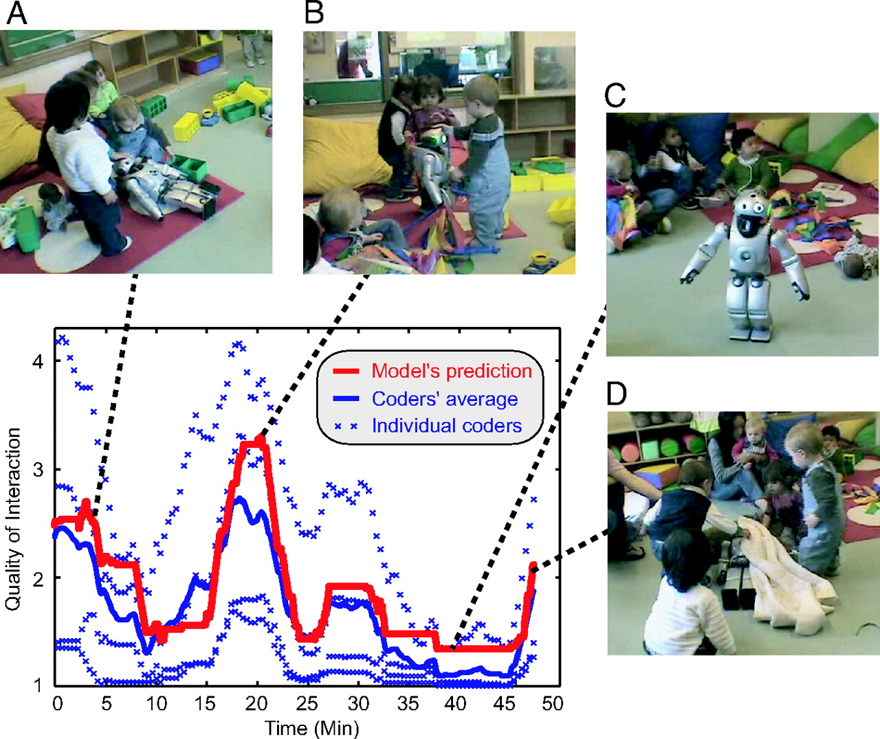
The Nov. 16 issue of Science has a special section on Robotics. I thought this graphic from the article by Pfeifer et al. - "Self-Organization, Embodiment, and Biologically Inspired Robotics" - was fascinating. It describes several biologically inspired robots.


Figure: Self-organization, dynamics, and materials in bio-inspired robotics. (A) Smooth transition between swimming and walking. This amphibious salamanderlike robot (~80 cm long) embeds a spinal cord model that explains the ability of salamanders to switch between swimming and walking. The locomotion model is built by extending a primitive neural circuit for swimming by phylogenetically more recent limb oscillatory centers. (B) Rich sensory stimulation through proper sensor morphology. This robot (~7 cm in diameter) owes its sophisticated sensory capacities to the specific arrangement, shape, and material characteristics of its whiskers. Natural whiskers from rodents (such as the ones used on this robot) are far superior to whiskers built from other materials in terms of richness of the signals relayed to the neural system. (C) Self-stabilizing rapid hexapod locomotion. This robot (~15 cm long) moves with a bouncing gait, achieving rapid (over 4 body lengths per second) locomotion. Its legs are built with compliant pneumatic actuators, which yield self-stabilization through mechanical feedback. (D) Passive dynamics–based walking. Designed to work on a slope as a dynamic walker, this robot (~45 cm tall) exploits dynamics and morphology (in particular, the shape and length of the body and feet) to achieve stable walking. The robot's natural dynamics serves as the target dynamics for a reinforcement learning mechanism, enabling the robot to quickly learn to walk on flat ground. (E) Self-stabilizing vertical takeoff through materials and morphology. Inspired by flies, this ultralight (60 mg, 3-cm wingspan) ornithopter (a device that flies by flapping its wings) generates sufficient lift to take off vertically (power is supplied externally). A large part of the control is delegated to the morphological and material properties of the robot. Compliant structures are driven into resonance to produce a large wing stroke, and flexible material is used in the wing hinges to allow for passive rotations of the wings. (F) Agile wall-climbing through materials. The bio-inspiration for this palm-sized robot is provided by the gecko and its uncanny climbing talents. The robot's tri-foot (three-footed wheel) is equipped with a polymer dry adhesive material, which to some extent has contact properties comparable to those of its biological analog. The robot can flexibly navigate on smooth vertical and even inverted surfaces. (G) Morphing through localized self-reconfiguration. This self-reconfigurable robot is composed of active (actuated, black) and passive (nonactuated, white) cubic modules (~400 g, ~60 to 65 mm side length). The modules connect to each other through hooks, which enables the robot to change its morphology in a large number of ways. The picture shows the metamorphosis from a four-legged (quadruped) structure to a linear (snakelike) structure. (H) Global movement through local interaction dynamics. The individual wheel-like modules (~10 cm in diameter) constituting this robot are equipped with spokelike parts driven by linear actuators. The wheels lie horizontally on the ground and attach to neighboring modules by Velcro. Although no module can move on its own, by using neural oscillators as drivers for the actuators and through the physical coupling between the units, a coordinated global wave of activation can be induced in clusters of more than 30 modules, which leads to forward movement, even though there is no global control.

Want to reduce risky behavior? Just pass a tiny current across your forehead.

Another brave new world item (from Fecteau et al.) - just what you need for your juvenile delinquent teenager! All you have to do is hook up a battery to two saline-soaked surface sponge electrodes to deliver anodal transcranial direct current stimulation (tDCS - two milliamps) to the right dorsolateral prefrontal cortex (DLPFC) to slightly up-regulate its activity, coupled with cathodal tDCS to the left DLPFC (current flows from anode to cathode). Here is the whole abstract and one figure:
Studies have shown increased risk taking in healthy individuals after low-frequency repetitive transcranial magnetic stimulation, known to transiently suppress cortical excitability, over the right dorsolateral prefrontal cortex (DLPFC). It appears, therefore, plausible that differential modulation of DLPFC activity, increasing the right while decreasing the left, might lead to decreased risk taking, which could hold clinical relevance as excessively risky decision making is observed in clinical populations leading to deleterious consequences. The goal of the present study was to investigate whether risk-taking behaviors could be decreased using concurrent anodal transcranial direct current stimulation (tDCS) of the right DLPFC, which allows upregulation of brain activity, with cathodal tDCS of the left DLPCF, which downregulates activity. Thirty-six healthy volunteers performed the risk task while they received either anodal over the right with cathodal over the left DLPFC, anodal over the left with cathodal over the right DLPFC, or sham stimulation. We hypothesized that right anodal/left cathodal would decrease risk-taking behavior compared with left anodal/right cathodal or sham stimulation. As predicted, during right anodal/left cathodal stimulation over the DLPFC, participants chose more often the safe prospect compared with the other groups. Moreover, these participants appeared to be insensitive to the reward associated with the prospects. These findings support the notion that the interhemispheric balance of activity across the DLPFCs is critical in decision-making behaviors. Most importantly, the observed suppression of risky behaviors suggests that populations with boundless risk-taking behaviors leading to negative real-life consequences, such as individuals with addiction, might benefit from such neuromodulation-based approaches.

Figure 1. Schematic representation of the experimental design. Each participant started to perform the risk task after receiving 5 min of stimulation. Stimulation continued throughout the task. The risk task was a decision-making task involving gambling. The task provided a measure of decision making under risk with little requirements on strategy and working memory.

Most popular consciousness articles...

For October 2007, from the ASSC archives (from which 49638 papers were downloaded from 69 countries!).
1. Rosenthal, David (2007) Consciousness and its function. In: 11th annual
meeting of the Association for the Scientific Study of Consciousness, 22-25
June 2007, Las Vegas, USA.
http://eprints.assc.caltech.edu/293/
2. Koriat, A. (2006) Metacognition and Consciousness. In: Cambridge handbook
of consciousness. Cambridge University Press, New York, USA. http://eprints.assc.caltech.edu/175/
3. Sagiv, Noam and Ward, Jamie (2006) Crossmodal interactions: lessons from
synesthesia. In: Visual Perception, Part 2. Progress in Brain Research,
Volume 155.
http://eprints.assc.caltech.edu/224/
4. Gennaro, Rocco J. (2007) Visual Agnosia and Higher-Order Thought Theory.
In: 11th annual meeting of the Association for the Scientific Study of
Consciousness, 22-25 Jun 2007, Las Vegas, USA. http://eprints.assc.caltech.edu/310/
5. Mashour, George A. (2007) Inverse Zombies, Anesthesia Awareness, and the
Hard Problem of Unconsciousness. In: 11th Annual Meeting of the ASSC, Las
Vegas. http://eprints.assc.caltech.edu/294/

Thursday, November 22, 2007

Predicting election outcomes in 100 milliseconds!

Another example of a quick judgment turning out to be more accurate than a considered one... Ballew and Todorov showed study participants transient pictures of the winner and runner-up for recent United States gubernatorial elections. Rapid, unreflective judgments of competence based solely on facial appearance (of candidates participants did not recognize) predicted the actual outcomes of gubernatorial elections. Instructions to deliberate and make a good judgment led to less accurate predictions of the election outcomes. Here is their abstract :
Here we show that rapid judgments of competence based solely on the facial appearance of candidates predicted the outcomes of gubernatorial elections, the most important elections in the United States next to the presidential elections. In all experiments, participants were presented with the faces of the winner and the runner-up and asked to decide who is more competent. To ensure that competence judgments were based solely on facial appearance and not on prior person knowledge, judgments for races in which the participant recognized any of the faces were excluded from all analyses. Predictions were as accurate after a 100-ms exposure to the faces of the winner and the runner-up as exposure after 250 ms and unlimited time exposure. Asking participants to deliberate and make a good judgment dramatically increased the response times and reduced the predictive accuracy of judgments relative to both judgments made after 250 ms of exposure to the faces and judgments made within a response deadline of 2 s. Finally, competence judgments collected before the elections in 2006 predicted 68.6% of the gubernatorial races and 72.4% of the Senate races. These effects were independent of the incumbency status of the candidates. The findings suggest that rapid, unreflective judgments of competence from faces can affect voting decisions.

Figure - An example of an experimental trial in the 250-ms presentation condition. Participants decided who was more competent.

Socialization between toddlers and robots

How's this for a (horrific) vision of the future! The abstract from Tanaka et al., followed by one figure from the paper:
A state-of-the-art social robot was immersed in a classroom of toddlers for >5 months. The quality of the interaction between children and robots improved steadily for 27 sessions, quickly deteriorated for 15 sessions when the robot was reprogrammed to behave in a predictable manner, and improved in the last three sessions when the robot displayed again its full behavioral repertoire. Initially, the children treated the robot very differently than the way they treated each other. By the last sessions, 5 months later, they treated the robot as a peer rather than as a toy. Results indicate that current robot technology is surprisingly close to achieving autonomous bonding and socialization with human toddlers for sustained periods of time and that it could have great potential in educational settings assisting teachers and enriching the classroom environment.


Figure - Predicting the quality of interaction. The red line indicates an automatic assessment of the quality of interaction between children and QRIO based on haptic (i.e. touching or physical contact) sensing. Blue lines indicate human assessment (by four independent coders) of the quality of interaction by using the continuous audience response method. (A) A session begins with QRIO waking up, attracting the children's interest. (B) During the music time in the classroom, children play with the robot. (C) Children are getting tired of the music time and losing interest in the robot. (D) Children put a blanket on the robot after it has laid down on the floor preparing for the end of a session.

Wednesday, November 21, 2007

Observing rat brains as they look to the future

An emerging view is that the hippocampus is essential to imagining the future as well as remembering the past (which makes a lot sense, since we usually base our imagined future on our past experience). Johnson and Redich have now observed ensembles of cells in the CA3 region of the rat hippocampus whose firing transiently encodes paths forward of an animal at decision points in a maze, as if they are reflecting on possible futures and deciding what to do next. The figure, from Heyman's review of the work in Science, illustrates that as a rat looks in one direction, neurons representing that position (inset) fire over a half-second period. Here is the abstract of the work:
Neural ensembles were recorded from the CA3 region of rats running on T-based decision tasks. Examination of neural representations of space at fast time scales revealed a transient but repeatable phenomenon as rats made a decision: the location reconstructed from the neural ensemble swept forward, first down one path and then the other. Estimated representations were coherent and preferentially swept ahead of the animal rather than behind the animal, implying it represented future possibilities rather than recently traveled paths. Similar phenomena occurred at other important decisions (such as in recovery from an error). Local field potentials from these sites contained pronounced theta and gamma frequencies, but no sharp wave frequencies. Forward-shifted spatial representations were influenced by task demands and experience. These data suggest that the hippocampus does not represent space as a passive computation, but rather that hippocampal spatial processing is an active process likely regulated by cognitive mechanisms.

Japanese Gardens

This picture from a stroll yesterday in the Morikami Japanese Gardens in Boca Raton, Florida. An iguana is on the lawn to the right of the tree trunk, a heron to its right on the bank, the flat object on the grass to the right of the heron is a water turtle, and the orange spots in the water are Koi. (click on the picture to enlarge).

Tuesday, November 20, 2007

Our optimism bias - brain correlates

Sharot et al. examine the tendency of most of us to imagine more optimistic outcomes than can be justified by sober appraisal. A moderate amount of optimistic illusion has been related to mental and physical health, and the general idea is that it is adaptive and useful because it motivates us towards future goals. Their abstract, and a figure showing the relevant brain regions:
Humans expect positive events in the future even when there is no evidence to support such expectations. For example, people expect to live longer and be healthier than average, they underestimate their likelihood of getting a divorce, and overestimate their prospects for success on the job market. We examined how the brain generates this pervasive optimism bias. Here we report that this tendency was related specifically to enhanced activation in the amygdala and in the rostral anterior cingulate cortex when imagining positive future events relative to negative ones, suggesting a key role for areas involved in monitoring emotional salience in mediating the optimism bias. These are the same regions that show irregularities in depression, which has been related to pessimism. Across individuals, activity in the rostral anterior cingulate cortex was correlated with trait optimism. The current study highlights how the brain may generate the tendency to engage in the projection of positive future events, suggesting that the effective integration and regulation of emotional and autobiographical information supports the projection of positive future events in healthy individuals, and is related to optimism.

The authors collected functional magnetic resonance imaging (fMRI) data while participants thought of autobiographical events related to a description of a life episode (for example, 'winning an award' or 'the end of a romantic relationship'). The word 'past' or 'future' indicated if they should think of an event that occurred in the past or one that might occur in the future. Trials were classified into positive, negative and neutral according to participants' ratings. They found that future positive events were rated as more positive than past positive events, and were imagined to be closer in temporal proximity then future negative events and all past events. (Click on image to enlarge it)

Impairment of action chains in autism.

When we observe the start of an action sequence that can end in two possible ways (in the figure shown a piece of food is placed in the mouth or in a container on the shoulder) appropriate sympathetic muscle EMG signals are detected at the start of the sequence. Thus, if the sequence will end in food to the mouth, activity is observed in the mouth-opening mylohyoid (MH) muscle at the onset. Rizzolatti and collaborators find that typically developing children show an activation of their MH muscle already when they observe the experimenter's initial motor act, food reaching. This activation reflects their understanding of the final goal of the observed action. In children with autism this action-understanding motor activation is lacking. Further, when typically developing children actually perform the observed action, MH muscle activation is observed at the very beginning of the sequence, while in children with autism, the activation is not observed until immediately before the muscle is actually used.

Figure - Schematic representation of the tasks. (Upper) The individual reaches for a piece of food located on a touch-sensitive plate, grasps it, brings it to the mouth, and finally eats it. (Lower) The individual reaches for a piece of a paper located on the same plate, grasps it, and puts into a container placed on the shoulder.

They suggest that high-functioning autistic children may understand the intentions of others cognitively but lack the mechanism for understanding them experientially because they lack the chains of action-constrained neurons that code specific motor acts (e.g., grasping) according to the final goal of the action in which the motor act is embedded.

Monday, November 19, 2007

Distinguishing true versus illusory memories with brain imaging.

Kim and Cabeza show that true versus illusory memories held with high certainty depend on different neural mechanisms. Here is their abstract and one figure from the paper:
Although memory confidence and accuracy tend to be positively correlated, people sometimes remember with high confidence events that never happened. How can confidence correlate with accuracy but apply also to illusory memories? One possible explanation is that high confidence in veridical versus illusory memories depends on different neural mechanisms. The present study investigated this possibility using functional magnetic resonance imaging and a modified version of the Deese-Roediger-McDermott false-memory paradigm. Participants read short lists of categorized words, and brain activity was measured while they performed a recognition test with confidence rating. The study yielded three main findings. First, compared with low-confidence responses, high-confidence responses were associated with medial temporal lobe (MTL) activity in the case of true recognition but with frontoparietal activity in the case of false recognition. Second, these regions showed significant confidence-by-veridicality interactions. Finally, only MTL regions showed greater activity for high-confidence true recognition than for high-confidence false recognition, and only frontoparietal regions showed greater activity for high-confidence false recognition than for high-confidence true recognition. These findings indicate that confidence in true recognition is mediated primarily by a recollection-related MTL mechanism, whereas confidence in false recognition reflects mainly a familiarity-related frontoparietal mechanism. This account is consistent with the fuzzy trace theory of false recognition. Correlation analyses revealed that MTL and frontoparietal regions play complementary roles during episodic retrieval. In sum, the present study shows that when one focuses exclusively on high-confidence responses, the neural correlates of true and false memory are clearly different.

Figure: Activity within medial temporal lobes (A) was greater for high-confidence true recognition (HC-TR) than for high-confidence false recognition (HC-FR). Activity within a frontoparietal network (B) was greater for high-confidence false recognition than for high-confidence true recognition.